

4.6.1 The digital microscope
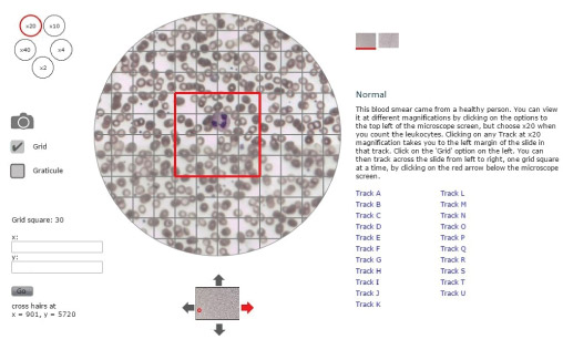
In the following activity, you will use the digital microscope to count leukocytes in microscope slides of blood smears that look like Figure 6. This figure shows a magnified blood sample from a healthy person (termed the ‘Normal’ sample). A second slide (not shown here) has blood from someone whose immune system was believed to be fighting a severe infection when the sample was taken. We have labelled this one ‘leukocytosis’ [loo-koh-sigh-toh-siss]. Leukocytosis refers to an increase in the total number of leukocytes of all types circulating in the bloodstream.
When you have counted the leukocytes in your two blood samples, you can compare your counts and decide whether there is evidence to suggest that the sample labelled ‘leukocytosis’ was taken from an individual who was indeed suffering from an infection.
Note the following features in Figure 6, which will help you to navigate the digital microscope when you access it online:
- You can switch between the ‘Normal’ and the ‘Leukocytosis’ slides by clicking on the squares to the top right of the microscope screen.
- The small circles to the top left of the microscope screen show the magnification factors you can choose for viewing the blood smears. They range from ×2 (twice the actual size of the cells) to ×40. Click on the circle labelled ×20 to choose the optimum magnification for this activity.
- When you click on the small square labelled ‘Grid’ on the left of the microscope screen, a matrix of faint grey lines is superimposed on the blood smear with a large red square in the centre of the grid. Your task is to count any leukocytes in the red square as you move it across the blood smear.
- The slides have been divided into horizontal sections labelled Track A to Track U, which are listed on the right of the microscope screen. It is not necessary to count all tracks on a slide although you can do if you wish. We suggest you just choose one track as a representative sample of the whole slide. When you click on a track (e.g. Track A) with the magnification set at ×20, the microscope screen shifts automatically to the left margin of the slide in that track.
- Notice the up/down/left/right arrows around the small image of the slide below the main microscope screen. If you click on the red arrow, it moves the slide to the right by one red grid square. This enables you to move across the blood smear one red grid square at a time until you reach the other side of the slide.