

1.4 Histology of the gut
The next type of tissue that we’ll examine more closely is that which forms the gut. Although the shape and dimensions of the gut vary along its length, the organisation of the different cell types that make up the gut wall is essentially similar throughout.
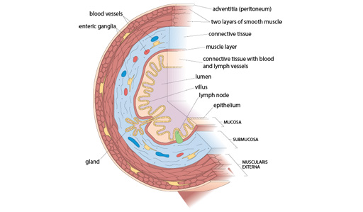
There are three main layers in the gut wall, as shown in Figure 1 below. Starting from the inside of the wall and working outwards these layers are the:
- mucosa
- submucosa
- muscularis externa.
Each of these layers, in turn, contains several cell types. Note that the adventitia (the name for the outer covering of any organ) does not form an integral part of the gut wall.
Let’s consider the composition of the three main layers of the gut wall in more detail.
Mucosa
In some areas such as the mouth, oesophagus and anus, where some physical protection is needed to prevent infection, the epithelium is several layers thick, and is something like that of the skin.
However, in most parts of the gut, the properties of the epithelium are rather different from those of the skin. Here the epithelium is only a single cell thick, with the apical membrane of these cells exposed to the lumen of the gut and the basal membrane facing the submucosal cells. All epithelial cells have two faces – the basal membrane lies on the connective tissue of the basal lamina and the apical membrane faces the surface of the tissue or duct.
These epithelial cells are specialised to perform digestive or absorptive roles. For example, the epithelial cells in the small intestine have small hair-like projections called microvilli on their apical surface.
Finally, in the stomach and intestines the epithelium is frequently invaginated (i.e. folded inwards) to form glands which consist of groups of secretory epithelial cells. Different types of gland are found in different regions of the gut.
Submucosa
The submucosa is a loose matrix of connective tissue in which blood and lymph vessels lie, and a large number of neuron cell bodies are grouped together as ganglia.
The enteric neurons (i.e. those associated with the gut) extend to the mucosa, where they influence both the production of digestive secretions by the epithelial cells and the state of dilation of the mucosal blood vessels.
Muscularis externa
The muscularis externa is a thick layer of smooth muscle. In almost all parts of the gut it consists of two separate muscle layers that lie at right angles to each other.
- The outer layer is longitudinal, that is it runs lengthways along the gut
- The inner muscle layer fibres encircle the wall of the tube.
Successive contraction and relaxation of these muscle layers results in peristalsis, which are waves of movement that propel the food through the gut.
Familiarise yourself with the overall anatomy of the gut before you move on to the next activity.