

1 Infection and inflammation
Histological examination of tissues can help diagnose disease, because each condition produces a characteristic set of changes in the tissue structure.
There are such a wide variety of diseases that histology alone usually cannot produce a diagnosis, although in some cases the histological appearance is definitive.
For example, a pathologist might see signs of a viral infection in the brain because of tissue damage and inflammation, but they would be unable to tell what virus is responsible for these changes. To identify the virus might require immunohistochemistry (IHC) for a specific viral protein or, more likely, the diagnosis would be confirmed by other symptoms and/or examination of a blood sample (serology).
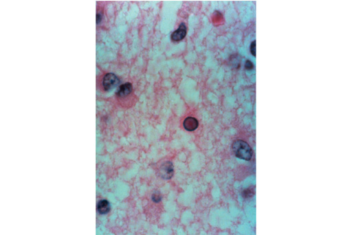
In some cases the histological appearance is characteristic of a specific disease. For example, the appearance of ‘owl-eye’ cells in the brain, as shown in the micrograph below, is diagnostic of a particular type of measles infection. However, normally histopathology reports only form one part of the picture of a disease that the clinician is trying to assemble.
Although diseases are very diverse, the responses made by the body are more limited, and fall into specific categories. For example, inflammation may be seen in response to an infection, as a result of physical damage or as part of an autoimmune disease, where the immune system attacks components of the body.
You’ll start looking at pathology in more detail by looking at two common causes of infection.