2.1 Cell death in the CNS
Cell death and degeneration within the nervous system is referred to as neurodegeneration. These processes occur in a number of genetic conditions as well as conditions associated with aging including Alzheimer’s disease and Parkinson’s disease.
Cell loss occurs in many tissues with age but the effects are particularly notable in tissues that have a limited capacity for regeneration, such as nerves in the CNS, the retina of the eye and the sensory cells of the inner ear. Histologically, it is more difficult to identify something that is not there than a change in the structure of the tissues.
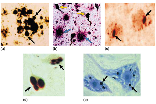
In diseases such as Alzheimer’s disease there is progressive loss of neurons and shrinkage of the brain, which may be more evident in the gross pathology, although counting the relative numbers of cells within an area can also give some histological indication of the cell loss. More evident are characteristic accumulations of proteins. Degenerating neurons leave tangles of fibres (neurofibrillary tangles) produced by degenerating components of the cytoskeleton. In addition, there are extracellular accumulations of amyloid within the brain.
Amyloid
Amyloid is an extracellular insoluble deposit of protein, and amyloidosis refers to the diseases in which amyloid occurs. In some cases production of amyloid is a primary event, and in others it is secondary to infection or a tumour. The actual protein type varies, depending on the cause of the condition. In some cases it affects individual organs, such as the brain in Alzheimer’s disease, but in the so-called ‘systemic amyloidoses’ many organs may be affected, including the lungs, kidney, heart and spleen.
There are a number of hereditary conditions in which the person lacks enzymes that break down particular macromolecules; they are collectively called storage diseases because the components that cannot be degraded within lysosomes accumulate and form insoluble deposits. This is particularly noticeable in the brain where they are often associated with neurodegenerative diseases; examples of this are shown in the micrographs below.