
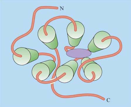
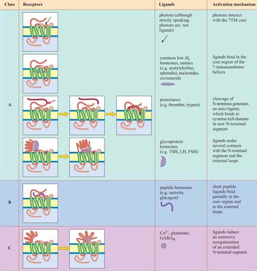
2.3.2 Seven-helix transmembrane (7TM) receptors
Although in unicellular organisms such as the yeast S. cerevisiae there are only two classes of 7TM receptors, the pheromone and glucose receptors, multicellular organisms have many more, accounting for up to 5% of all genes in C. elegans and 2% of genes in the human and Drosophila genomes. 7TM proteins have been classified into four classes, A, B, C (Table 1). Between them, they can bind a huge range of ligands including simple ions, nucleotides, lipids, steroids, modified amino acids, peptides and glycoprotein hormones at a variety of binding sites, and even photons can activate certain 7TM receptors. An example of the binding of a ligand (adrenalin) to its 7TM receptor is shown in Figure 22. As a result of this wide range of ligands, the mechanisms of activation of 7TM receptors are also extremely diverse. They may include, for example, proteolytic cleavage of the N-terminus, and subsequent binding of the new peptide fragment to the central core or ligand binding to the N-terminus or to the central core. However, in all cases the end result is a change in the conformation of the 7TM receptor (Table 1).
Can you recall any examples of 7TM receptors?
Muscarinic cholinergic receptors and adrenergic receptors.
*Activation of the A, B and C classes of 7TM receptors involves coupling to G proteins
In the case of GPCRs, ligand binding influences the equilibrium between the active and inactive conformation of the receptor in favour of the active conformation, altering the interaction of the cytosolic loops of the protein with a trimeric G protein (which may be already associated with the receptor in a preformed complex). In turn, this brings about activation of the G protein (described in Section 3.2).
What is the fundamental difference between signalling through nicotinic and muscarinic ACh receptors?
Nicotinic receptors do not employ a signal transduction pathway to effect their action: the binding of the ligand directly opens a Na+ion channel linked to the receptor, and causes membrane depolarization, resulting in the contraction of skeletal muscle cells. Muscarinic ACh receptors, being GPCRs, activate signal transduction pathways via G proteins.