3.4 Specialisation within language areas: brain scanning
Is there any evidence from the undamaged brain that the view derived from aphasia is indeed correct? The most useful methodologies here use either PET or functional MRI (fMRI) scanning to establish which parts of the brain are active in particular tasks. The difficulty is that a standard linguistic task, such as understanding a sentence's meaning, involves phonology and syntax and semantics, and thus is not helpful when trying to tease out which of these subtasks happens in which areas.
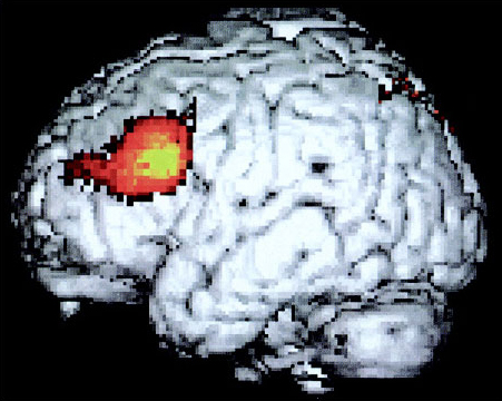
Many studies have looked at the pattern of activation produced in the brain by single words. The areas especially active are widespread and somewhat variable, but generally include the auditory cortex on both sides, other parts of the left temporal lobe, and Wernicke's area. A study by Karin Stromswold from the Massachusetts Institute of Technology aimed to identify the areas specialised for the processing of syntax (Stromswold et al., 1996). Her team set up two different conditions of sentence processing. In one condition the participants heard sentences like The child spilled the juice that stained the rug, whereas in the other they heard sentences like The juice that the child spilled stained the rug. Both of these contain the same words. The first is syntactically quite simple because the order of the nouns in the sentence mirrors their logical relations (child spilled juice, juice stained rug). The second is more complex as the order of its elements does not reflect the logical relations. The areas of the brain specialised for syntax should be more active in the second condition than the first.
The most significant difference between the first and second conditions was indeed that Broca's area was much more active in the second (Figure 17). This finding confirms that of several other studies. Thus the view from aphasia seems confirmed; the anterior language areas are specialised for syntax (and verbs and sentence construction), whereas the posterior and temporal ones are more specialised for individual word meanings (and nouns and concreteness). This is doubtless a simplification. There is evidence of significant variability between individuals, and the distinction between areas and subtasks is not watertight. Moreover, we usually do all the subparts of linguistic processing interactively and simultaneously, so something that affects any one part will probably affect them all to a greater or lesser extent. Nonetheless, research in this area is allowing us to understand the anatomy of the language faculty in greater and greater detail.