2 X-ray imaging
2.1 Introduction
X-ray imaging is probably the best known and most widely used of the imaging techniques we will cover. Not only are X-rays the longest established means of producing images of the internal structure of the body, but X-ray imaging is also the workhorse technique for all radiology departments.
X-rays are high-energy electromagnetic radiation; those used for diagnostic imaging typically have photon energies between 20 and 120 keV.
In X-ray imaging, a beam of X-rays is passed through the patient and detected on the opposite side. As the X-rays pass through the body some are either absorbed or scattered. This attenuation depends on the thickness of the material and on its attenuation coefficient. The equation that describes the reduction in intensity is

where I0 and I are the intensity before and after passing through material of thickness x and μ is the attenuation coefficient.
Bone has a much higher attenuation coefficient than soft tissue, which in turn has a higher coefficient than air. This means that, if a patient is placed in a uniform beam of X-rays, radiation that has passed through soft tissue will have a higher intensity at the detector than radiation that has passed through bone. Hence the image will show good contrast between bone and soft tissue and, similarly, between soft tissue and lungs. X-ray images usually show the high attenuation material (e.g. bone) as white and the low attenuation material as black.
Watch the video clip below which shows Alan having X-rays of his skull taken by a radiographer, following his arrival by ambulance to the Accident and Emergency Department with a possible head injury and skull fracture.
Click to view the clip about planar x-ray images [2 minutes 45 seconds]
Transcript: Planar x-ray images
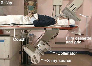
Figure 3 shows the X-ray unit used to image Alan's skull. It's main components – the x-ray source, collimator, patient couch, and film and cassette holder – are labelled.