
2.2 Components of an X-ray unit
2.2.1 X-ray source
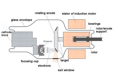
X-rays are produced when energetic electrons strike a metal target. The X-ray source consists of an evacuated tube containing a cathode, from which the electrons are emitted, and an anode, which supports the target material where the X-rays are produced. Only about 1 per cent of the energy used is emitted as X-rays – the remainder is dissipated as heat in the anode. In most systems the anode is rotated so that the electrons strike only a small portion at any one time and the rest of the anode can cool. The X-rays are emitted from the tube via a radio-translucent exit window.
Some of the X-rays are given off at an energy that depends on the nature of the target material. These are known as characteristic radiation (see Figure 5). There is also a broad spectrum of radiation known as bremsstrahlung (braking radiation); it is this radiation that is used in most diagnostic procedures (see Figure 5). However, both types contribute to the radiation dose given to the patient.
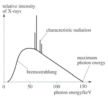
The maximum photon energy is determined by the voltage applied to the tube (known as kVp). The peak of the X-ray spectrum is at about half of this energy. kVp values vary between about 20 kV for very thin body parts and 120 kV for a pelvic X-ray. Filters can be placed in front of the exit window to eliminate low-energy X-rays that do not contribute to the final image.