
3 Computed tomography
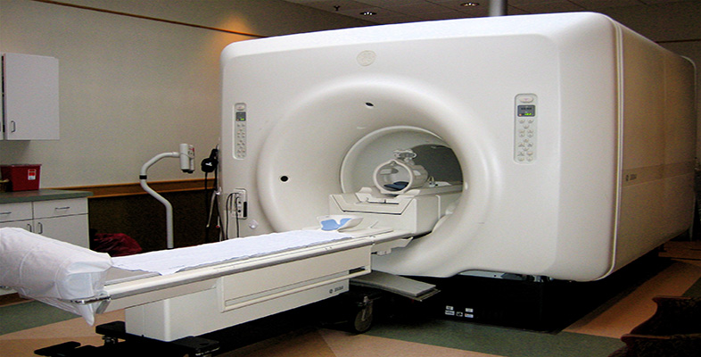
The aim of computed tomography (CT) is to produce an image of a slice of the body. (The Greek word ‘tomos’ means slice.) This is achieved by rotating a thin, fan-shaped beam of X-rays around the patient and measuring the intensity on the opposite side of the patient with a very large number of detectors.
The following video clip shows Alan having a CT scan of his head to see if he has a base of skull fracture. Listen and watch the video clip carefully, with the following questions in mind:
Activity 5
What are the main differences between the skull X-rays and CT images produced of Alan?
What colour do the skull bone and brain tissue appear on the CT images and why?
How does the video say a two dimensional ‘slice’ is produced through the body?
Click to view the clip about computed tomography [2 minutes 47 seconds]
Transcript: Computed Tomography (CT)
Answer
The skull X-rays showed ‘projections’ of Alan's skull (a 3D object represented in 2D) and the brain tissue could not be seen. The CT slices show the skull and internal brain tissue as a series of ‘slices’, and therefore, in much more detail.
The skull bone appears white and the brain tissue grey. This is because the skull attenuates X-rays to a greater extent than the brain tissue, and just like a planar X-ray the more a tissue attenuates X-rays, the whiter it will appear in the final image.
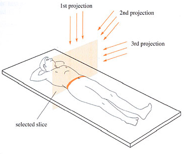
The X-ray source is rotated around the patient and the intensity recorded on the opposite side of the patient. Using data from a large number of angles a computer reconstruction can produce a two-dimensional map of the tissues in a slice of the body.
Let's look at the answer to the final question in more detail. First we will consider exactly how the X-ray source rotates around the patient.
In modern scanners the source and the detectors rotate around the patient at more than one revolution per second. In the older scanners the couch was moved after one rotation and the subsequent rotation had to be in the opposite direction to avoid twisting the cables (called ‘stop and go’ by Dr Klaus Klingenbeck in the following video clip). However, with the introduction of slip-rings it became possible to keep the source and detectors rotating continuously and to move the couch at the same time. This means that the X-ray source describes a helix around the patient and a set of data covering the complete volume of the patient can be collected. This is known as spiral (or helical) scanning and has the advantage that the data for the entire thorax or other section of the body can be collected in one breath hold. More recently, multislice scanners, in which there is more than one arc of detectors, allow even faster data collection.
Click to view part 1 of the clip about X-rays and CT [2 minutes 1 second]
Transcript: X-rays and CT - part 1
Now let's consider how a ‘slice’ through the patient can be reconstructed by the transmission data obtained at a large number of different angles, using a technique called ‘filtered back projection’. The following video clip will look at the transmission data from simple objects and how back projection, and finally filtered back projection, are used to reconstruct the original object from the transmission data. This clip introduces some complex topics – you only need a general overview of how CT image reconstructions work.
Click to view part 2 of the clip about X-rays and CT [8 minutes 36 seconds]
Transcript: X-rays and CT - part 2
Activity 6
Now that you understand more about the CT imaging equipment and the images it produces, take a little time to consider what you think the advantages and disadvantages of this technique would be.
Answer
The advantages of CT imaging are:
excellent resolution and contrast;
choice of tomographic or 3D-images available;
relatively fast (compared with MRI);
contrast medium can be used.
The disadvantages are:
larger dose of ionizing radiation than most planar X-ray procedures;
equipment is costly and therefore not available at all hospitals;
slower and more complex to undertake than most planar X-ray procedures.