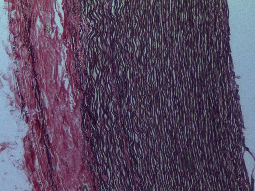
Figure 5 Section across the wall of the human aorta stained with elastic Van Gieson, which identifies the elastic fibres (black) in the media of the artery and the collagen (red) in the adventitia.

Personalise your OpenLearn profile, save your favourite content and get recognition for your learning

Start this free course now. Just create an account and sign in. Enrol and complete the course for a free statement of participation or digital badge if available.