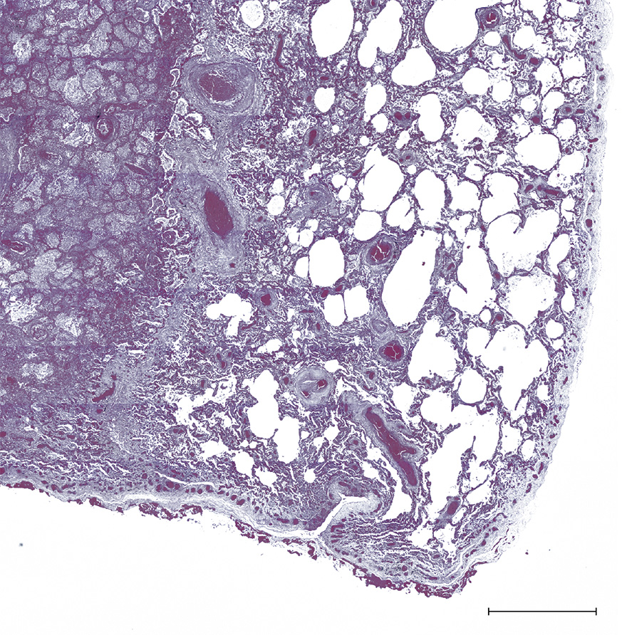
Figure 6 An area of necrosis in the lung caused by blockage of the blood supply is visible on the left of this section. The area on the right includes surviving lung tissue, which is thickened and has areas of fibrosis. Scale bar = 2 mm.

Personalise your OpenLearn profile, save your favourite content and get recognition for your learning

Start this free course now. Just create an account and sign in. Enrol and complete the course for a free statement of participation or digital badge if available.