

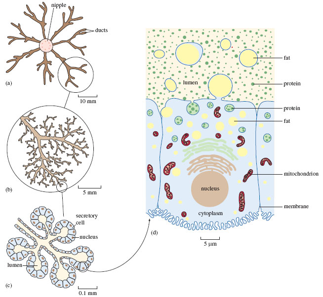
Figure 17 The structure of a mature mammary gland, at increasing magnification: (a)-(c) viewed under a light microscope, (d) with an electron microscope. (a) and (b) show the arrangement of the milk-carrying ducts and (c) the secretory cells. (d) is an image of a single cell, showing the structures involved in the processes of secretion. The different scales show the extent of magnification. (1 mm, i.e. one millimetre, is equivalent to 1000 μm, i.e. 1000 micrometres, and 0.1 mm = 100 μm)