
3.2 Colour vision
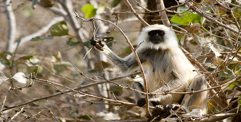
DA stresses that colour vision is very important in primates, not only because colour is used 'in sexual displays' such as advertising a female's receptiveness to mating [p. 275], but also to identify ripe fruit [p.247] and to select nutritious leaves [p. 255]. This section discusses these points in more detail and explains how the visual system in primates is able to detect colour.
White light is composed of light of different wavelengths, from 300-800 nanometres (nm); 1 nm is one millionth of a millimetre. To understand colour vision you don't need to know anything about the physical properties of light, other than each band of wavelengths produces light of a particular colour (see Figure 1).
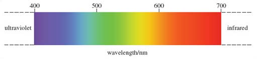
You may be familiar with the terms used to describe the shortest and the longest wavelengths, namely ultraviolet and infrared - over-exposure to ultraviolet light produces sunburn. Between these two extremes lie the wavelengths of the so-called visible spectrum, the light of various colours that humans can detect.
Light is detected by the retina at the back of the eye. In primates, as in the majority of mammals, light is detected by two types of so-called photoreceptor cell, namely rods and cones, which contain special light- sensitive compounds called photopigments. As stated by DA, cones are 'those elements in the retina that detect colour'.
So rods and cones are the receptor cells of the retina, and they are directly linked to nerve cells - more technically termed neurons. These neurons transmit information (in the form of tiny electrical signals) via the optic nerve to the visual cortex, the region of the brain that processes visual information. All neurons are interconnected; indeed, those with a specialised linking function are termed interneurons. They receive signals from one or more neurons and pass them to others, forming neural networks between the brain and the body. Each neuron has a long extension called an axon, which enables it to transmit a signal over a considerable distance before passing it to another neuron via a junction termed a synapse.
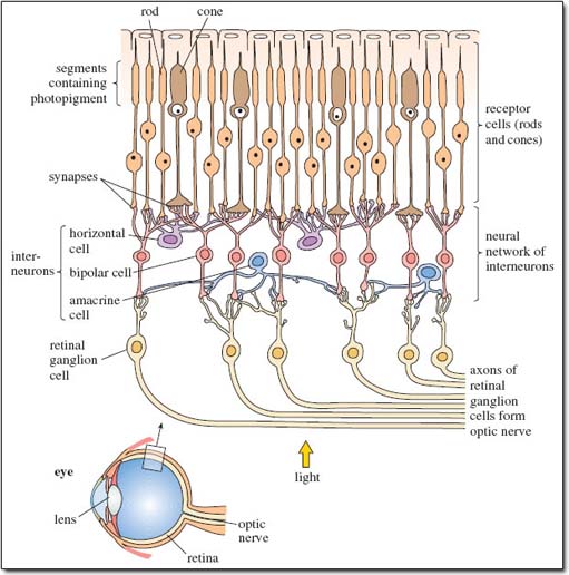
Figure 2 is a stylised diagram of the neural network of the retina showing the connections between the various cells. Spend a little time studying it. Locate the light-sensitive rods and cones (towards the top of the diagram) and the neural network of interneurons beneath them, which you'll appreciate is more complex in reality than shown here. Locate synapses, axons and various types of interneuron.
Light falling on the rods and cones produces signals that pass from receptor cells to interneurons, termed bipolar cells, and then to retinal ganglion cells whose axons form the optic nerve. There are two more types of interneuron in this network - locate them on Figure 2 - that form connections between different groups of rods and cones, such that signals from several receptor cells contribute to each signal entering the optic nerve. The communications that occur between these interneurons are too complex for discussion here, but this rich network contributes significantly to the sensitivity of primate vision. Rods are able to respond to low levels of light because a large number of adjacent rods (between 15 and 45) are linked to a single interneuron. Light only has to fall onto one of the rods for a signal to be transmitted to the brain. Not surprisingly, rods are the main or in some cases, as DA mentions on p. 257, the only component of the retina of nocturnal primates and are important for night vision in prosimians.
Cones need higher levels of light than rods to be activated and each cone is connected to between one and four neurons. In most species of New World monkeys, and indeed in all other non-primate mammals, cones contain two photopigments, one that responds to short-wavelength light and one that responds to medium-wavelength light; these photopigments may be found in the same cone or in separate cones. Electrical input from the activated cones is combined to produce colour images. This type of vision is termed dichromatic, in recognition of the two pigments.
Question 4
Dichromatic vision is not full colour vision, the type of vision that humans share with other anthropoid primates. Suggest why this is.
Answer
Because, unlike us, these animals cannot detect longer-wavelength light - they cannot distinguish red from other colours. (Refer back to Figure 1.)
Activity 2
You will recall from LoM and the TV programme that the only nocturnal anthropoids are owl monkeys. Watch the TV programme from 15.20-16.39 and reread LoM p. 257. Write a few sentences explaining why owl monkeys are thought to be descended from diurnal ancestors and how they compensate for poor vision.
Answer
Owl monkeys, like other nocturnal primates, have large eyes with retinas that contain many rods but no cones. Prosimians, however, also possess a tapetum lucidum, which reflects light towards the retina, greatly enhancing vision in low light. The lack of a tapetum in owl monkeys suggests that ancestral owl monkeys were diurnal, as are all other anthropoid species. In compensation for their poor vision, owl monkeys have an improved sense of smell compared with primates possessing colour vision. They mark trails through the branches, using a scent-impregnated tuft of hair that lies just beneath the tail.
How have you coped with the detailed anatomical diagram, Figure 2, showing the arrangement of cells in the retina at the back of the eye? Looking at the way different colours are used in the diagram should have helped you. You might find it useful to draw your own simplified version of Figure 2, perhaps as a series of boxes labelled 'rods and cones', 'interneurons', 'retinal ganglion cells' and 'optic nerve', linked by arrows to show the direction in which the nerve signals travel. Add some large arrows showing the direction of the light entering the eye - which, as the caption says, is not what you'd expect. Light reaching the rods and cones has had to travel through all the other cells. That gives you some idea of the transparency of cells, which are, in reality, packed very closely together, not arranged with lots of spaces between them as shown for clarity in this simplified figure.