

1 Identifying cell types within tissues
This is a short introduction to some of the types of cells that you will encounter when you start to examine tissues under the microscope.
Cells in the body differentiate into many types as they develop, and within a single organ there may be many different types of cell. For example, in the liver the major cell type is the hepatocyte, which carries out many metabolic functions, but there are also cells associated with blood vessels, bile ducts and immune defence, as well as various structural cells.
Despite the diversity of cells there are a number of patterns of cell organisation which occur in many different tissues. Some key examples are described in the text below.
Epithelial cells
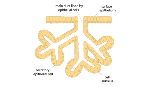
Epithelial cells form tight cohesive sheets of cells which cover many of the body surfaces, such as the skin or the gut. They also form secretory glands, such as the breast, tear ducts, sweat glands and salivary glands. The ducts leading from the secretory cells in these tissues are lined with other types of epithelium.
A layer of epithelial cells may be just a single cell thick, or may be several cells deep. This difference affects their overall shape, which may be cuboidal, columnar or stratified (flattened). You can see some of this variation in the diagram below.
Endothelial cells
Another type of cell that is found in layers is the endothelial cell. These form a single layer that lines blood or lymphatic vessels.
Muscle cells
Muscle cells fall into three main types.
- Striated muscle fibres form the voluntary muscles of the body, i.e. the ones that you can choose to move.
- Smooth muscle is a contractile tissue found in many tissues which operates involuntarily. It is found in the gut and around blood vessels and many glands and ducts.
- Cardiac muscle is a specialised muscle found only in the heart.
Nerve cells (neurons)
Neurons have long processes (axons) which extend away from the nerve body. They form both the:
- central nervous system (CNS), namely the brain, spinal cord and retina of the eye
- the peripheral nervous system, which describes any nervous tissue outside the CNS.
Nerves run through virtually all tissues of the body, and there are large networks of nerves in tissues such as the gut.
Adipose tissue (adipocytes)
This type of cell stores fat, and is found in many tissues, not just in the main fat storage areas of the body. Adipocytes have a large central vacuole where lipids (fats) are stored.
Leukocytes (white blood cells)
As you read and observed in Week 1, leukocytes come in a number of different types. They are found in all tissues of the body (not just the blood), where they carry out the function of immune defence.
Lymphoid tissues, such as lymph nodes, tonsils, adenoids and Peyer’s patches (a lymphoid tissue of the intestine), contain highly organised groupings of leukocytes that activate and coordinate the body’s immune response to infection.
Extracellular matrix proteins
Bone, tendons and connective tissues are mostly formed of extracellular matrix proteins which are produced by specific cell types within the tissue. For example, collagen is a major extracellular matrix protein, and it is formed in several different types, depending on its function within the tissue.