Skip to content
Skip to main content

About this free course

Download this course

Share this free course

Understanding autism

Start this free course now. Just create an account and sign in. Enrol and complete the course for a free statement of participation or digital badge if available.

4.1 Brain structure and function

Research into the structure and function of the brain draws extensively on a range of brain imaging techniques. Magnetic Resonance Imaging (MRI) suggests that key brain structures may have a slightly different size or shape in autistic people. For instance, studies suggest that the brains of some young autistic children are 5–10% bigger than those of typically developing children, although this difference disappears by adolescence. Another area where increased size has been observed is the amygdala, a brain region involved in evaluating the emotional significance of external events. Overgrowth of the amygdala in children with autism is related to the severity of their social and communication difficulties, – greater overgrowth tends to go with more severe difficulties – but again this disparity of size compared with typical development disappears in adolescence.

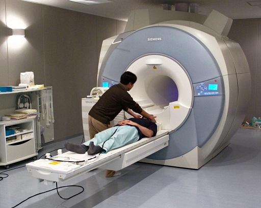
Functional Magnetic Resonance Imaging (fMRI) monitors brain activity while a person is performing psychological tests, such as recognising faces, responding to emotional stimuli or understanding language.

A photograph shows a person about to be scanned by an fMRI machine.
Figure 6 Image of Functional Magnetic Resonance Imaging (fMRI) scanning. The participant in an fMRI study responds to images, sounds or other stimuli while lying in a scanner. Use of magnetic fields to monitor blood flow in the brain yields information about which brain regions are active.

The patterns of brain activity revealed by fMRI may differ in people with autism, compared to the neurotypical population. For instance, there may be reduced activity in a brain region called the fusiform gyrus, which has a specialised role in face recognition, linking with the observation that autistic people find it hard to recognise faces which they have seen before.

Two side-by-side fMRI images show the right and left cerebral cortex, and the cerebellum.
Figure 7 Images of fMRI scans of an adolescent male on the autism spectrum (right) compared with an age- and IQ-matched typically developing control (left).

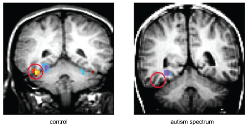
Atypical patterns of brain activity are also observed when autistic people perform tasks such as the ‘Reading the Mind in the Eyes’ test illustrated earlier.

(See Lai, Lombardo and Baron-Cohen, 2013, for an overview of findings like those discussed in this section.)