
Immunoelectron microscopy
Immunolabelling to localise specific molecules to particular cell organelles can also be applied in electron microscopy. In EM immunolabelling (often known as immunoelectron microscopy, or immuno-EM), the antibody molecules are tagged with electron-dense substances, the most effective being small gold particles, which can be observed as dark dots. Different antibodies can be tagged with particles of different sizes, allowing detection of more than one type of molecule to be performed simultaneously.
Figure 3 shows an example of how immunocytochemistry at the EM level can provide information about the localisation of molecules in an individual cell. The figure shows two cells of a bacterium, Neisseria meningitidis, labelled with an antibody that recognises a particular oligosaccharide (short chain of sugar units). The binding of this primary antibody is visualised using a secondary antibody coupled to gold particles, 15 nm in diameter, each of which can be seen as a very dark (electron-dense) dot.
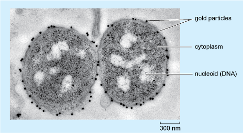
Looking at Figure 3, in which part of the bacterium is the oligosaccharide located?
The gold particles are, with only one exception, found on the outer surface of the two bacteria; so it can be deduced that this is where the oligosaccharide is predominantly localised in N. meningitidis.