Microtubules
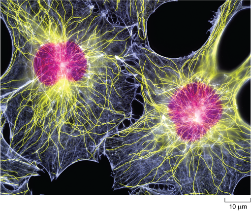
Microtubules are, as their name implies, tubular structures composed of two forms (called α and β) of a protein known as tubulin. They are present in all eukaryotic cells and play a crucial role in maintaining cell shape and also in intracellular movement: for example, the movement of cell organelles from one part of the cell to another, and the reorganisation of chromosomes into the daughter cells during cell division. Microtubules are effectively hollow tubes typically consisting of 13 parallel filaments of tubulin assemblies, and they measure about 25 nm in external diameter (Figure 8b).
In animal cells, microtubules radiate from a microtubule organising centre (MTOC) known as the centrosome which is located near the nucleus (Figure 1a and Figure 9). Typical plant cells do not have an equivalent of the centrosome. Instead, less well-defined microtubule organising centres (MTOCs) appear at different times and places to organise microtubule networks in different types of plant cell. Microtubules are very unstable, and are constantly being disassembled and reassembled. They begin to assemble at the centrosome, and the growing tubules extend out radially, towards the edges of the cell (Figure 10). Some microtubules disassemble before they reach the cell cortex, but they will become stabilised if they attach to an organelle or bind to a protein known as a capping protein.
What might happen to the shape of a cell if the capping proteins become confined to one part of the cell cortex?
Microtubules would only become stabilised at that region of the cell periphery, so the cell could become polarised; that is, it could acquire an asymmetrical shape.
What types of animal cell are inherently asymmetrical in shape?
Neurons and some kinds of epithelial cells are examples of asymmetrical cells .
The selective stabilisation of microtubules, determined by the location of capping proteins, is therefore an important factor in determining cell shape. The arrangement of microtubules also plays an important role in the distribution of organelles within the cell.