
2.1 Computed tomography (CT) scans
A conventional X-ray of a bone fracture is a two-dimensional (2D) image, taken from the front of the patient by a single camera, and is sometimes known as a planar X-ray.
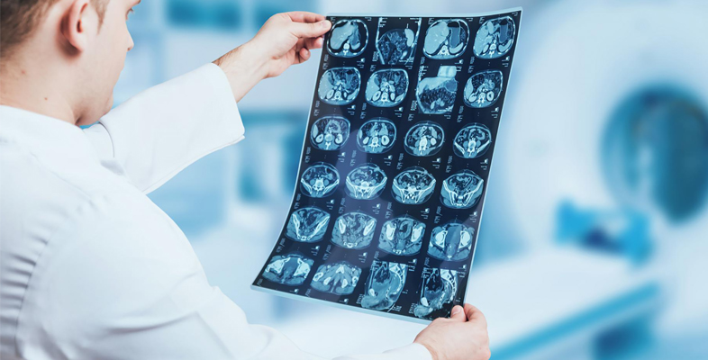
However, a computed tomography (CT) scan produces many 2D images of sections throughout the body using detectors arranged in a circular field, which can then be computer processed to give a three-dimensional (3D) reconstruction of the body.
With carefully controlled conditions, even changes in soft tissues indicating tumours can be picked up and located by this method. The resolution can be as good as 1 mm or less.
The following video shows a CT scan being done in a hospital for a patient with a suspected injury to his spine.
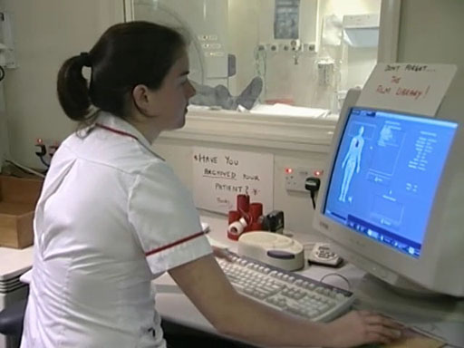
Transcript: Video 1 X-ray imaging in a CT scan. (2:32 min)
RADIOGRAPHER: Okay, I’m going to take some pictures of your head now, okay? So what I’m going to do is just going to raise the bed up and get you into position ...
NARRATOR: Alan is now being placed in the CT scanner, and the radiographer uses a laser beam to place his head in precisely the right location. CT scanning is essentially an X-ray procedure, but the X-ray source is rotated around the patient and the intensity recorded on the opposite side of the patient.
Using data from a large number of angles, a computer reconstruction can produce a two-dimensional map of the tissues in a slice of the body.
RADIOGRAPHER: So what we’re going to do now is choose the protocols that we’re going to use to do the head. So we’re going to click on the appropriate part of the body here. So we click on ‘head’. And we’re going to do an adult brain.
Okay, the scan’s just about to start, so just keep nice and still there, okay?
NARRATOR: Planning is done by taking a pilot scan. The patient is moved through the gantry. The source remains stationary on one side. This produces an image similar to a planar X-ray. This pilot scan is taken in what is called the sagittal plane.
RADIOGRAPHER: We’re going to do thinner sections through the base of the skull, and then we’re going to do wider ones as we go through towards the head at the top of the skull there. So now we can do our actual scan, okay? And then we can produce the actual images that the radiologist will look at.
So we can just scroll through the images that we’ve just got, okay? And we can alter the grey levels of the images so we can see different bits of anatomy.
What we’ll do is we’ll look at the actual brain windows first. So we’re going to go through, okay? We’re looking basically for any asymmetry or any abnormalities.
When we get further up, we can change the window levels, so we can see the ventricles better. Just going through there. Now we’re at the top.
And as this patient has got suspected basal skull fracture, what we’d now do is go back and we’re going to change it to bony windows. We’ve highlighted – the bone is white and everything else is now dark – so it’s not seen so clearly.
So again, we’re just looking through to see if there’s any dark lines in the skull which could represent fractures.
NARRATOR: The concern over the potential for injury to the top of the spine has been dismissed. But there is a concern that there may be some damage to the brain. So it’s decided to do an MRI scan.
So, how is a CT image produced?
The X-ray source is rotated around the patient and the intensity recorded on the opposite side of the patient. Using data from a large number of angles, a computer generates a two-dimensional map of the tissues in a slice of the body.
Note that there are three directions in which slices through the brain (or the body in general) are typically reported in imaging, as illustrated in Figure 3: axial, sagittal and coronal. You’ll meet these terms again when looking at MRI.
