

5.3 Platinum binding to DNA
Oligonucleotides are short sections of DNA (2 to 15 nucleotides long) used for model studies. They are often referred to as a ‘duplex’ in recognition of the two-stranded helical structure of DNA.
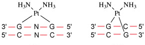
The early structural characterisation of oligonucleotide-bound cisplatin was done using X-ray crystallography, and molecular modelling. Cisplatin was found to form a cis complex with two adjacent guanines on the same strand (known as a G(N7)–p–G(N7) linkage, often shortened to G–p–G or G–G). This is shown in Figure 19.
Experiments found that cisplatin formed several different types of adduct with the DNA oligomers.
The major product (60–65%) is the G–G 1,2-intrastrand link between guanine residues which reside in the major groove in the B-form DNA (also shown in Figure 20): cis-[Pt(NH3)2(G–p–G)] (Structure 6).
Smaller quantities of other adducts include:
- 20–25% of a G–A 1,2-intrastrand: cis-[Pt(NH3)2(A–p–G)], (Structure 7).
 Structure 6 (left) and 7 (right)
 - 5–10% of more widely spaced guanine adducts: the 1,3-intrastrand and G–p–G interstrand complexes (Structure 8, 9 and Figure 20). (Note: N in Structure 8 represents another base.) In the latter, cisplatin forms a cross-link between the two strands.
 Structure 8 (left) and 9 (right)
- 2–5% of a monoadduct (Structure 10), and
 Structure 10 (left) and 11 (right)