
5.4 Anticancer effect
There is evidence that cisplatin induces cells to undergo apoptosis (programmed cell death).
This occurs because the cell recognises the damaged DNA and triggers the mechanisms that signal the cell to die.
This self-destruct mechanism is present in all cells and is part of the organism’s way of destroying cells that might be harmful to itself.
See Box 2 for information on cell death and DNA repair systems.
Box 2 DNA repair systems and cytotoxicity: why do cells die?
Damage is constantly caused to cells and DNA by normal metabolism and by external factors such as UV radiation, smoking, chemicals, and so on. Sometimes this leads to the damaged cell swelling and then bursting – a form of cell death known as necrosis. DNA is continually checked for ‘errors’ in its sequence by various proteins.
If there is a large amount of damage, enzymes come into play that trigger the death of a cell by apoptosis or programmed death. If the damage is not too great, the checking proteins activate enzymes to eradicate the errors and perform a ‘repair’ by excising the damaged part and reconstituting the sequence. Indeed, it is the existence of these DNA repair systems that is believed to lead to resistance to cisplatin in certain cancers. On the other hand, studies have shown that repair-deficient mutant cells are much more sensitive to cisplatin.
Professor Lippard talks you through the effect of cisplatin on the autorepair mechanisms in the following video. You’ll also see the gel electrophoresis technique being used to separate DNA fragments.
Transcript: Video 10 The cisplatin story: Part 5. (3:42 min)
NARRATOR: Formation of the cisplatin DNA adduct has damaged the DNA. If the DNA is not repaired then the cell cannot reproduce itself or replicate. Research has moved on to examine the cell mechanisms that deal with this damage. Proteins are the key to processes such as the repair mechanism. Normally, damaged DNA is recognised and marked for repair, which is done by cutting out the damage, so-called excision repair.
The bent, platinated DNA seems to be recognised by other proteins which bind at the damaged site and prevent access by the repair proteins.
STEPHEN LIPPARD: The class of proteins that bind to the cisplatin-modified DNA, it turns out from gene sequencing and other experiments that we’ve done, generally contain what are called high-mobility group, or HMG, domains.
NARRATOR: The term ‘high-mobility group’ comes from this procedure known as electrophoresis. It’s used to identify and separate individual strands of DNA from a mix of all sorts. Cancer cells are cultured in the Petri dishes at body temperature, 37 degrees Celsius. The cells are harvested and the protein extracted.
They are then mixed with platinated DNA, which has also been made radioactive by including a radioactive isotope of phosphorus – 32 phosphorus – in the phosphate groups. The cultures are incubated for varying amounts of time and dyed a bright colour to make them visible.
The culture is dropped onto a gel and an electric field then applied. The DNA is negatively charged because of the phosphate groups. And so it begins to travel down the gel towards the positively charged cathode. To make a permanent record of the experiment, a photographic film is put next to the gel. The radioactive phosphorus isotope blackens the film, thus indicating the positions of the pieces of DNA.
As the repair mechanism operated during the incubation, bits of the DNA that are damaged by the cisplatin were excised. Because these pieces are smaller and lighter, they travel faster and further down the gel. The longer the incubation ran, the more small excised bits of platinum-containing DNA can be seen as the DNA tries to repair itself.
The technique can be used to study the effect of the HMG proteins on the repair mechanism.
STEPHEN LIPPARD: They could bind to the cisplatin-modified DNA and shield or protect the adduct from excision repair. If this happened more efficiently in a tumour cell than in a normal cell of the same tissue, that would be a wonderful mechanism for anticancer activity. And we’re working hard to evaluate that hypothesis.
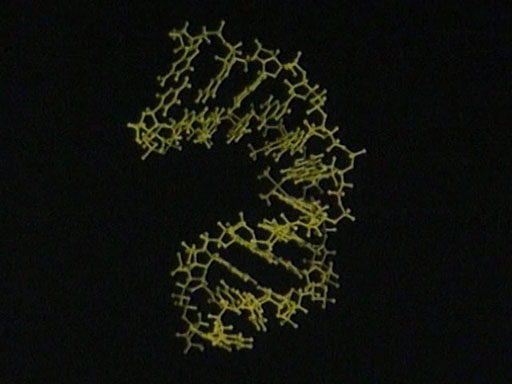
NARRATOR: This structure shows an HMG protein – the red tube – attached to a natural binding site for transcription. It binds to the minor groove. But the similarity of the effect on the minor groove to the effect of cisplatin is striking. In both cases, the minor groove becomes wider and shallower. Perhaps it is possible that the cisplatin DNA confuses the repair mechanism by this similarity and so escapes excision.
-
What type of protein is thought to bind to cisplatin-modified DNA, preventing access by repair proteins?
-
High-mobility-group (HMG) proteins.
So in general, cisplatin appears to inhibit repair in mutant cells, leading to cell death. The difficult question to be answered is how do Pt 1,2-intrastrand cross-links inhibit repair?
One hypothesis is that the binding of HMG protein HMGB1 with the 1,2-intrastrand cisplatin–DNA complex shields the DNA from intracellular repair, leading to apoptosis. This binding occurs by means of HMG inserting a phenyl group protruding from its backbone into the notch created when cisplatin forms a complex with DNA.
This also increases the bend in the DNA even more to about 60–90°, facilitating the binding of a signalling protein called P53 which triggers a cascade of events leading to cell death. However, there is equally compelling evidence suggesting that HMGB1 can cause these changes even in the absence of cisplatin-induced lesions. So it is clear that these complex mechanisms still remain an area where much research is required.
Figure 23 shows schematically how these mechanisms are thought to operate.