
When a murder has occurred, forensic science can be essential for proving the identity of both victim and killer. Some killers have wrongly assumed that the absence of a complete body, or the removal of distinguishing features such as fingerprints, would prevent the identification of a victim. The combination of different types of forensic science, in particular dentistry, pathology and entomology, can be used to establish the identity both a victim and their killer. Far from needing a complete body, analysis of only small remnants of a person, such as teeth or fragments of bone, can be used to confirm who a victim was. It is not so easy to dispose of a body, and that evidence is left behind even when a corpse is subject to fire or acid.
 DNA profiling (sometimes called DNA fingerprinting) can be used to establish the identity of a suspect. DNA is made from molecules called nucleotides that are joined together to produce a long chain. In fact, DNA is comprised of two of the long nucleotide chains that wrap around each other to form a helix. The discovery of the DNA helix in 1953 by Francis Crick and James Watson was a critical breakthrough in understanding the genetic inheritance. For their discovery, Crick and Watson (and their colleague Maurice Wilkins) were awarded the 1962 Nobel Prize in Physiology or Medicine. The nucleotides within the DNA chain have a chemical component called a nucleobase. There are four different nucleobases that make up DNA, They are adenine, thymine, guanosine and cytosine (usually denoted by the letters A, T, G and C). The order in which these nucleobases occur along the length of a DNA strand essentially determines the physical characteristics of an individual.
DNA profiling uses techniques that can reveal small variations in the sequence of nucleobases from one person to another. DNA profiling techniques have become a powerful resource for modern forensic science, and have even allowed old, unsolved cases to be reviewed with fresh evidence. When a person dies their DNA will begin to degrade, but this can take a long time. In fact, some tissues can provide DNA samples even after a body has been buried for some time. Bone and dental pulp, for example, are sometimes used as a source of DNA for profiling when bodies are exhumed. DNA profiling is not used by itself to establish guilt, but can be used in conjunction with other evidence to implicate a person in a crime, or exonerate them.
The first step in DNA profiling is the collection of a DNA sample. This is done using strict procedures and storage methods. DNA can be obtained from many different sources, including hairs, blood stains, semen, urine, and skin cells in sweat or saliva. Typically, the DNA is purified from the sample and separated from other cellular contents using solvents or digestive enzymes that remove proteins, fats and other unwanted materials. Once purified, the concentration of the DNA sample is measured so scientific tests can use known amounts.
 Alec Jeffreys invented the DNA profiling technique in 1984
The purified DNA sample can be used in a number of different ways. At present, a common approach is to look at DNA regions that have sequences called short tandem repeats. A short tandem repeat is a sequence of DNA that can be 2 or more nucleotides in length, and is repeated many times over, with one repeat occurring just next to another. The number of times that a particular short tandem repeat occurs is characteristic of an individual’s DNA sequence. So by analysing the length of short tandem repeat sequences, it is possible to establish the likelihood that a DNA sample came from a particular person.
Alec Jeffreys invented the DNA profiling technique in 1984
The purified DNA sample can be used in a number of different ways. At present, a common approach is to look at DNA regions that have sequences called short tandem repeats. A short tandem repeat is a sequence of DNA that can be 2 or more nucleotides in length, and is repeated many times over, with one repeat occurring just next to another. The number of times that a particular short tandem repeat occurs is characteristic of an individual’s DNA sequence. So by analysing the length of short tandem repeat sequences, it is possible to establish the likelihood that a DNA sample came from a particular person.
For DNA profiling to give useful results, it is important to examine the short tandem repeats occurring in many designated DNA regions. For example, the UK uses 17 DNA regions (16 DNA regions with short tandem repeat sequences and a DNA region that identifies gender; referred to as DNA-17). The value of analysing short tandem repeats in many regions of DNA is that it provides a strong statistical case for identifying who a DNA sample may belong to. It is considered that there is only a one in a one-billion chance of two individuals having the same patterns of short tandem repeats within the DNA regions. The only exception to this is identical siblings, which share that the same DNA sequences.
To be able to analyse the short tandem repeats within in DNA sample, it is necessary to copy the DNA using a technique called the polymerase chain reaction (usually known by the acronym PCR). This technique was developed by the Nobel Prize-winning scientist Kary Mullis in 1983, and is a relatively simple method for amplifying only specific parts of a DNA sequence. Mullis demonstrated that amplification of specific DNA sequences could be achieved by using a pair of short nucleotide chains known as primers. The primers are designed to bind solely to the nucleobase sequences within the DNA region of interest.
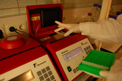 A scientist placing a strip of eight PCR tubes, each containing a reaction mixture, into the thermal cycler
PCR is a cyclical process in which DNA is repeatedly heated and cooled, and new nucleotide chains are made through the action of an enzyme called DNA polymerase. The primers direct the DNA polymerase to amplify only the region of DNA where they bind. Using PCR it is possible to obtain many copies of a DNA sequence even if the sample obtained from a crime scene was minimal or somewhat degraded. It takes somewhere between 30 to 60 minutes to amplify a DNA sequence using PCR. The actual time depend on how many cycles are needed, and how good the starting template DNA material is. PCR amplifications are carried out using an automated machine called a thermal cycler, which as its name suggests is able to undertake the necessary cycles of heating and cooling.
A scientist placing a strip of eight PCR tubes, each containing a reaction mixture, into the thermal cycler
PCR is a cyclical process in which DNA is repeatedly heated and cooled, and new nucleotide chains are made through the action of an enzyme called DNA polymerase. The primers direct the DNA polymerase to amplify only the region of DNA where they bind. Using PCR it is possible to obtain many copies of a DNA sequence even if the sample obtained from a crime scene was minimal or somewhat degraded. It takes somewhere between 30 to 60 minutes to amplify a DNA sequence using PCR. The actual time depend on how many cycles are needed, and how good the starting template DNA material is. PCR amplifications are carried out using an automated machine called a thermal cycler, which as its name suggests is able to undertake the necessary cycles of heating and cooling.
Once a PCR procedure has finished, and a specific DNA sequence has been amplified, the results have to be analysed. There are several ways of doing this. A common method is to use a technique called gel electrophoresis in which the DNA sample from the PCR procedure is introduced into a jelly-like matrix called a gel. Once the DNA sample is loaded into the gel a voltage is applied from the top to bottom of the gel, with the negatively-charged terminal (cathode) at the top and the positively-charged terminal (anode) at the bottom. Gel electrophoresis works because the nucleotides that make up DNA contain phosphate, which is negatively charged. The negatively-charged phosphate means that DNA molecules will migrate in the direction of positive charge when the voltage is switched on.
DNA samples are usually added near to the top of the gel, so that they can migrate down towards the anode. The gel matrix acts as a kind of a sieve that serves to retard the DNA as it migrates. A long strand of DNA will migrate more slowly than a short strand because it is retarded more. Gel electrophoresis therefore allows DNA molecules to be accurately separated depending on their size. By separating DNA in this way it is possible to reveal whether a DNA sample came from a particular individual. A simple illustration of how this could be done is shown in the figure below.

The figure shows a hypothetical situation in which 3 PCR procedures were performed using different template DNA. One DNA sample was obtained from a tissue sample at the crime scene. The other DNA samples were obtained from individuals. The DNA samples were used for PCR to amplify specific sequences, and the product of the PCR procedure was subject to gel electrophoresis. The DNA sequences that were amplified can be seen as discrete bands on the gel. The bands represent the same-sized DNA sequences that were amplified many times over during the PCR procedure. By comparing the bands, it is possible to determine whether the original DNA samples are matching. For example, in the figure it appears that the DNA sample from the crime scene is a match for the DNA sample from individual 2 because the same bands were amplified during the highly specific PCR procedure.
Explore more...
-
Forensic science and fingerprints
Learn more to access more details of Forensic science and fingerprintsThis free course, Forensic science and fingerprints, covers how science can make fingerprints easier to study, how they are used in court and some of the questions about the extent to which fingerprint identification is sound and scientific. Students will learn the principles used in classifying and matching fingerprints (often called marks).
Level: 1 Introductory
Rate and Review
Rate this article
Review this article
Log into OpenLearn to leave reviews and join in the conversation.
Article reviews