Use 'Print preview' to check the number of pages and printer settings.
Print functionality varies between browsers.
Printable page generated Saturday, 11 July 2026, 7:08 PM
Antenatal Care Module: 7. Physiological Changes During Pregnancy
Study Session 7 Physiological Changes During Pregnancy
Introduction
During pregnancy, a woman’s body changes in many ways due to the effect of hormones. These changes can sometimes be uncomfortable, but most of the time they are normal and enable her to nourish and protect the fetus, prepare her body for labour, and develop her breasts for the production of milk.
Can you recall the definition of a hormone from Study Session 3?
Hormones are signalling chemicals produced in the body, which circulate in the blood. Different hormones control or regulate the activity of different cells or organs.
In this study session, you will learn about some of the changes that occur during pregnancy in the uterus, cervix and vagina, the cardiovascular system, gastrointestinal system, and urinary system, and about changes in the breasts and skin. You will also learn about the implications of all these changes for you as a health worker managing the health of pregnant women. By understanding the normal changes of pregnancy, you can reassure the woman if she is concerned, and also detect and intervene more quickly if you notice any abnormalities. A basic knowledge of these changes and adaptations is also critical for understanding the results of laboratory tests that may be conducted at a health facility during the pregnancy.
Learning Outcomes for Study Session 7
When you have studied this session, you should be able to:
7.1 Define and use correctly all of the key words printed in bold. (SAQ 7.6)
7.2 Describe physiological changes in the female reproductive system during pregnancy and the consequences of these changes for the pregnant woman. (SAQs 7.1 and 7.6)
7.3 Describe the average changes in the pregnant woman’s body weight. (SAQ 7.2)
7.4 Discuss changes in the cardiovascular system during pregnancy, and the effects on blood pressure, cardiac output, blood volume and red blood cell concentration. (SAQs 7.3, 7.4 and 7.6)
7.5 Recognise normal and abnormal changes in the pregnant woman’s respiration, digestion, urinary system, skin and breasts, including the production of colostrum. (SAQs 7.5 and 7.6)
7.1 Physiological changes in the female reproductive system during pregnancy
7.1.1 Changes in oestrogen and progesterone
In Study Sessions 3, 4 and 5 you learned about the main female reproductive hormones, oestrogen and progesterone, and their functions in preparing the uterus for pregnancy. Oestrogen and progesterone are also the chief hormones throughout pregnancy.
A woman will produce more oestrogen during one pregnancy than throughout her entire life when not pregnant. During pregnancy, oestrogen promotes maternal blood flow within the uterus and the placenta.
How does oestrogen play an important role in the development of the fetus?
By promoting maternal blood flow to the uterus and placenta it ensures that the fetus is supplied with nutrients and oxygen for its development, and that waste products from the fetus are removed in the mother’s blood. (You learned about this in Study Session 5.)
A pregnant woman’s progesterone levels are also very high. Among other effects, high levels of progesterone cause some internal structures to increase in size, including the uterus, enabling it to accommodate a full-term baby. It has other effects on the blood vessels and joints, which we will discuss later in this study session.
7.1.2 Changes in the uterus, cervix and vagina
The uterus
After conception, the uterus provides a nutritive and protective environment in which the fetus will grow and develop. It increases from the size of a small pear in its non-pregnant state to accommodate a full-term baby at 40 weeks of gestation. The tissues from which the uterus is made continue to grow for the first 20 weeks, and it increases in weight from about 50 to 1,000 gm (grams). After this time, it doesn’t get any heavier, but it stretches to accommodate the growing baby, placenta and amniotic fluid. By the time the pregnancy has reached full term, the uterus will have increased to about five times its normal size:
- In height (top to bottom) from 7.5 to 30 cm
- In width (side to side) from 5 to 23 cm
- In depth (front to back) from 2.5 to 20 cm.
What causes these changes?
The hormone progesterone is primarily responsible.
At 12 weeks’ gestation (near the end of the first trimester, i.e. three-month period), the fundus (upper margin of the body of the uterus) may be palpated (felt) through the abdomen above the pubic bone (symphysis pubis). The size of the uterus usually reaches its peak at about 36 weeks’ gestation (see Figure 7.1).
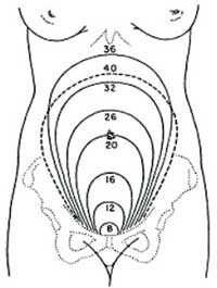
After how many weeks of pregnancy can the fundus be palpated at the height of the woman’s umbilicus (belly-button), according to Figure 7.1?
At about 20 weeks.
The uterus may drop slightly as the fetal head settles into the pelvis, preparing for delivery. Notice the position at 40 weeks of gestation, which is shown as a dotted line in Figure 7.1. This dropping is referred to as ‘lightening’. It is more noticeable in a primigravida (pregnant for the first time) than in a multigravida (a woman who has been pregnant previously, regardless of outcome).
The cervix
The cervix remains 2.5 cm long throughout pregnancy. In late pregnancy, softening of the cervix occurs in response to increasing painless contractions of its muscular walls.
The vagina
The vagina also becomes more elastic towards the end of pregnancy. These changes enable it to dilate during the second stage of labour, as the baby passes down the birth canal. (You will learn all about this in the next Module on Labour and Delivery Care.)
7.2 Pregnancy-related changes in posture and joints
A pregnant woman’s entire posture changes as the baby gets bigger. Her abdomen transforms from flat or concave (dished) to very convex (bulging outwards), increasing the curvature of her back. The weight of the fetus, the enlarged uterus, the placenta and the amniotic fluid (the bag of waters surrounding the baby), together with the increasing curvature of her back, puts a large strain on the woman’s bones and muscles. As a result, many pregnant women get back pain. Too much standing in one place or leaning forward can cause back pain, and so can hard physical work. Most kinds of back pain are normal in pregnancy, but it can also be a warning sign of a kidney infection. (You will learn how to identify kidney infections in Study Session 18.)
In addition, progesterone causes a loosening of ligaments and joints throughout the body. Pregnant women may be at greater risk of sprains and strains because the ligaments are looser, and because their posture has changed.
7.3 Changes in body weight during pregnancy

Continuing weight increase in pregnancy is considered to be one favourable indication of maternal adaptation and fetal growth. However, routine weighing of the mother during pregnancy is not now thought to be necessary, because it does not correlate well with pregnancy outcomes. For example, there can be a slight loss of weight during early pregnancy if the woman experiences much nausea and vomiting (often called ‘morning sickness’). You will learn more about this and other minor disorders of pregnancy in Study Session 12, later in this Module. The expected increase in weight of a healthy woman in an average pregnancy, where there is a single baby, is as follows:
- About 2.0 kg in total in the first 20 weeks
- Then approximately 0.5 kg per week until full term at 40 weeks
- A total of 9 -12 kg during the pregnancy.
A woman who is pregnant with more than one baby will have a higher weight gain than a woman with only one fetus. She will also require a higher calorie diet. (You will learn a lot about diet and healthy nutrition in pregnancy in Study Session 14 of this Module.)
A lack of significant weight gain may not be a cause for concern in some women, but it could be an indication that the fetus is not growing properly. Doctors and midwives may refer to this as intrauterine growth restriction (IUGR) of the fetus.
7.4 Changes in the cardiovascular system
The cardiovascular system consists of the heart, the blood vessels (veins and arteries), and the blood that circulates around the body. It is the transport system that supplies oxygen and nutritive substances absorbed from the gastrointestinal tract to all the cells, tissues and organs of the body, enabling them to generate the energy they need to perform their functions. It also returns carbon dioxide, the waste product of respiration, to the lungs, where it is breathed out. The chemical processes that go on in the body generate many waste products, which the blood transports to the kidneys and liver, where they are removed. Other functions of the cardiovascular system include the regulation of body temperature, and the circulation and delivery of hormones and other agents that regulate body functions. There are several significant changes in this complex system during pregnancy.
7.4.1 The heart
The heart may increase in size during pregnancy due to an increase in its workload.
Why do you think the workload of the heart has to increase?
Because it has to pump blood through the placenta, fetus and the much larger uterus and abdomen of the pregnant woman.
The amount of blood that is pumped out of the heart each minute is called the cardiac output. Table 7.1 shows how it increases during pregnancy.
| Woman’s condition | Cardiac output (litres per minute) |
|---|---|
| non-pregnant, resting | 2.5 |
| end of 1st trimester | 5 |
| end of 2nd trimester | 6 |
| full-term | 7 |
The increase in cardiac output is caused by two changes in how the heart functions:
- Increase in the resting heart rate, i.e. the number of heart beats per minute. The heart rate is about 15 beats per minute higher in the pregnant woman.
- Increase in the stroke volume, i.e. the volume of blood pumped out of the heart in a single heart beat. It is about 7 millilitres (ml) larger per heart beat in the pregnant woman.
Cardiac output is calculated by multiplying heart rate and stroke volume.
During the second trimester of pregnancy, the mother’s heart at rest is working 40% harder than in her non-pregnant state. Most of this increase results from a more efficiently performing heart, which ejects more blood at each beat.
7.4.2 Blood volume
Blood volume (the total volume of blood in the circulation, measured in litres) increases gradually by 30-50 % in the pregnant woman, so by full term she has about 1.5 litres more blood than before the pregnancy. A higher circulating blood volume is required to provide extra blood flow through the placenta, so nutrients and oxygen can be delivered to the fetus. The increase in blood volume is caused by two changes:
- Increase in the volume of blood plasma (the fluid part of the blood).
- Increase in the number of red blood cells in the circulation.
The volume of blood plasma increases after about the sixth week of pregnancy. It reaches its maximum level of approximately 50% above non-pregnant values by the second trimester, and maintains this until full term.
The total volume of red cells in the circulation increases by about 18% during pregnancy, in response to the extra oxygen requirements made by the maternal, placental and fetal tissues. Red blood cells contain the oxygen-carrying substance called haemoglobin, which is rich in iron (see Box 7.1). Taking iron supplements during pregnancy can result in a much greater increase in red blood cells, up to 30% more than non-pregnant levels.
Haemoglobin is pronounced ‘heem oh gloh binn’.
Box 7.1 Iron, haemoglobin and anaemia
Iron is present in all cells and has several important functions, including oxygen transport and storage in the human body. It is the critical component of the oxygen-carrying substance haemoglobin, found in all red blood cells. It is the iron in haemoglobin that makes these cells appear red. If the diet is too low in iron, the person cannot make enough red blood cells. Iron is also involved in the storage and release of oxygen in the muscles.
Anaemia is most accurately defined as a low concentration of haemoglobin in the blood, but it is often referred to as a low concentration of red blood cells. Too little iron in the diet is the leading cause of anaemia.
The measurement of haemoglobin is expressed using its chemical symbol (Hb), and its weight in grams (gm) per decilitre (dl) of blood. A decilitre equals 10 millilitres (ml). The World Health Organization recommends that the pregnant woman’s haemoglobin should not fall below 11 grams of haemoglobin per decilitre of blood (Hb 11g/dl). (You will learn more about anaemia and its treatment in Study Session 18.)
Although there is a constant increase in the number of red blood cells in the circulation during pregnancy, the increase in the volume of blood plasma is much larger. So even though the pregnant woman has more red blood cells than before she was pregnant, they are diluted in the much larger volume of blood plasma.
What effect will this have on the concentration of red blood cells and haemoglobin in the pregnant woman’s blood, compared to her non-pregnant self?
The concentration of red blood cells and haemoglobin will fall because they are more dilute, so the woman’s blood will be slightly anaemic.
This effect is referred to as physiological anaemia. It explains why iron in the diet, or from iron tablets, is so important during pregnancy.
7.4.3 Blood pressure in pregnancy
We said earlier that progesterone causes the ligaments and joints to loosen during pregnancy. It also acts with some other natural chemicals in the body to cause the muscular walls of the blood vessels to relax slightly. The result is that there is less resistance to the flow of blood around the body, because the same volume of blood is circulating in slightly wider blood vessels. Blood pressure (BP) refers to how hard the blood is ‘pushing’ on the walls of the major blood vessels as it is pumped around the body by the heart.
What effect will the slight relaxation of the blood vessel walls have on the pregnant woman’s blood pressure?
It will be lower than in her pre-pregnant state, because the same volume of blood has more space in which to circulate.
Lower blood pressure is particularly common in early pregnancy. Many women report occasionally feeling dizzy in the first trimester, because less blood and less oxygen is being pumped to the brain. Progesterone can also cause a sudden larger relaxation in the blood vessels, resulting in an acute feeling of dizziness, or even a brief loss of consciousness (passing out).
Another cause of dizziness can result from lying flat on the back. This is more common after 24 weeks of pregnancy, but it can happen earlier during twin pregnancies, or conditions that increase the volume of amniotic fluid (waters surrounding the fetus). When a pregnant woman is lying flat on her back, the weight of her uterus and its contents compresses the large blood vessel (vena cava) leading from her lower body to the heart. When this blood vessel is squashed, the blood flow back to the heart is reduced, which in turn leads to a reduction in the blood flow out of the heart to the rest of the body.
If there is suddenly less blood leaving the heart, what will happen to the woman’s blood pressure, and how will she feel as a result?
Lying flat on her back can result in a sudden and dramatic drop in blood pressure, and dizziness or loss of consciousness may occur because not enough oxygen is reaching her brain. After the first trimester, pregnant women are recommended not to lie on their back.
7.4.4 Exercise and blood flow in pregnancy
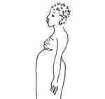
The weight gain in pregnant women increases the workload on the body from any physical activity. Steady, non-violent exercise is good for the mother because it prepares her body for labour (Figure 7.2), but sudden strong exercise, or working for too many hours without a break, may make her feel dizzy. This is because the reduced blood pressure and slight physiological anaemia cannot keep pace with the demand of her body for more oxygen.

A pregnant woman should not do exercises where she is lying on her back, due to the compression of the major blood vessels returning blood to her heart. Strong exercise may lead to decreased blood flow to the uterus because blood is diverted to the muscles, but this has not been shown to have any long-term effects on the baby. Pregnant women should not exercise vigorously in hot weather, or if it makes them breathless, or if there are known risk factors such as a history of miscarriage.
7.4.5 Oedema in pregnancy
Oedema is pronounced ‘ee dee mah’.
![]() If a pregnant woman experiences severe oedema, including swelling of the face, this is a danger sign that requires immediate referral to the nearest health facility.
If a pregnant woman experiences severe oedema, including swelling of the face, this is a danger sign that requires immediate referral to the nearest health facility.
A combination of the slight increase in the permeability of the smallest of blood vessels (they allow more fluid to leak out into the tissues), the additional weight of the uterus, and the downward force of gravity, slow down the rate at which blood is pumped back to the heart from the lower half of the body. Fluid often collects in the tissues of the legs and feet of pregnant women after the first trimester, instead of being absorbed into the blood circulation. The swelling caused by this collection of fluid is called oedema.
It is a common condition in pregnant women, particularly if they stand for a long time during the day. Oedema of the hands may also occur. Advise the woman to rest frequently and to elevate (raise) her feet and legs while sitting. This will improve the return of blood to her heart and decrease swelling of the legs.
7.5 Respiratory changes
During pregnancy, the amount of air moved in and out of the lungs increases by nearly 50% due to two factors:
- each breath contains a larger volume of air
- the rate of breathing (breaths per minute) increases slightly.
During pregnancy, many women find they get short of breath (cannot breathe as deeply as usual). This is because the growing baby crowds the mother’s lungs and she has less room to breathe. But if a woman is also weak and tired, or if she is short of breath all of the time, she should be checked for signs of sickness, heart problems, anaemia or poor diet. Get medical advice if you think she may have any of these problems.
7.6 Changes in the gastrointestinal system in pregnancy
As you may remember from your high school biology, food and fluids enter the gastrointestinal system in the mouth, pass through the oesophagus, stomach and intestines, and solid waste exits at the anus. This very long tube from mouth to anus is often called the ‘gut’. Proteins, fats and carbohydrates in our diet are broken down (digested) in the gut into units small enough to be absorbed from the intestines into nearby blood vessels. It is also the route by which nutritious substances, such as vitamins and minerals, enter the body.
During pregnancy, the muscles in the walls of the gastrointestinal system relax slightly, and the rate at which food is squeezed out of the stomach and along the intestines is slowed down.
Can you think of a reason why slowing down the passage of food through the gastrointestinal system might be beneficial in pregnancy?
It increases the time available for digestion, and it maximises the absorption of nutrients from the diet.
Undesirable effects also result from slow emptying of the stomach, and slow movement of food through the gut.
Can you suggest one of these undesirable effects?
Many pregnant women experience constipation (difficulty in passing stools).
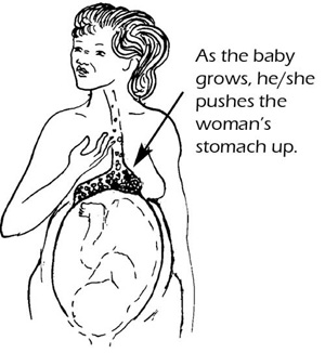
Many women also have nausea in the first months of pregnancy. A burning feeling, or pain in the stomach or between the breasts, is called indigestion (or ‘heartburn’, although the heart is not involved). It happens because as the pregnancy progresses, the growing baby crowds the mother’s stomach and pushes it higher than usual (Figure 7.3). The acids in the mother’s stomach that help digest food are pushed up into her chest, where they cause a burning feeling. This is not dangerous and usually goes away after the birth.
If the mother has difficulty with nausea or indigestion, advise her to eat small, frequent meals. The mother should not lie down flat for 1 to 2 hours after eating, because this may cause these symptoms. In Study Session 12 you will learn more about minor disorders of pregnancy such as these, and how to help the woman manage them.
7.7 Changes in the urinary system during pregnancy
The urinary system consists of the kidneys (a pair of organs on either side of the abdomen near the back), the tubes connecting the kidneys to the bladder where urine is stored, and a tube called the urethra that passes urine out of the body. (Look back at Figure 3.1 in Study Session 3, to remind yourself of the position of the bladder and the urethra.) The kidneys extract waste from the blood and turn it into urine. They must work extra hard to filter the mother’s own waste products from her blood, plus those of the fetus, and get rid of them in her urine. Therefore, there is also an increase in the amount of urine produced during pregnancy.

Needing to urinate (pee) often is normal, especially in the first and last months of pregnancy. This happens because the growing uterus presses against the bladder. In late pregnancy, a woman often has to get up during the night to urinate, because fluid retained in the legs and feet during the day (oedema) is absorbed into the blood circulation when her legs are raised in bed. The kidneys extract the excess fluid and turn it into urine, so the bladder fills more quickly at night.
7.8 Skin changes
Changes in the woman’s hormones, and mechanical stretching of her growing abdomen and breasts, are responsible for several changes in the skin during pregnancy.
7.8.1 Linea nigra
This dark line may appear between the umbilicus (belly-button) and the symphysis pubis (pubic bone); in some pregnant women it may extend as high as the sternum (the bone between the breasts). It is a hormone-induced excess production of brown material (pigment) in the skin cells in this area. After delivery, the line begins to fade, though it may never completely disappear.
7.8.2 Mask of pregnancy (chloasma)
Some women produce a brownish pigmentation of the skin over the face and forehead, known as the ‘mask of pregnancy’ (or chloasma). It gives a bronze look. It begins about the 16th week of pregnancy and gradually increases, but it usually fades after delivery. You will learn more about it in Study Session 8.
7.8.3 Stretch marks
As the woman’s weight increases, stretching of the skin occurs over areas of maximal growth — the abdomen, thighs and breasts. Pink or brownish stretch marks may appear in some women, which can be quite dramatic. They usually fade after delivery, although they never completely disappear.
7.8.4 Sweat glands
Activity of the sweat glands throughout the body usually increases during pregnancy, which causes the woman to perspire (sweat) more profusely than usual, particularly in hot weather or during physical work.
7.9 Changes in the breasts
In early pregnancy, the breasts may feel full or tingle, and they increase in size as pregnancy progresses. The areola around the nipples (the circle of pigmented skin) darkens and the diameter increases. The Montgomery’s glands (the tiny bumps in the areola) enlarge and tend to protrude (stick out more). The surface blood vessels of the breast may become visible due to increased circulation, and this may give a bluish tint to the breasts.

By the 16th week (during the second trimester), the breasts begin to produce colostrum. This is the precursor of breastmilk. It is a yellowish secretion from the nipples, which thickens as pregnancy progresses. It is extremely high in protein and contains antibodies (special proteins produced by the mother’s immune system) that help to protect the newborn baby from infection. Near the end of pregnancy, the nipples may produce enough colostrum to make wet patches on the woman’s clothes. Reassure her that this is normal and a good sign. After the baby is born, colostrum is produced for about the first three days, before the proper milk begins to flow. Make sure that the mother breastfeeds the colostrum to her baby, so he or she gets all the nutrients and antibodies it contains.
In Study Session 8, you will meet some signs of these physiological changes in pregnancy again, when you learn how to diagnose whether a woman is pregnant, and how to gather information about her ‘pregnancy history’.
Summary of Study Session 7
In Study Session 7, you have learned that:
- Oestrogen and progesterone are the chief pregnancy hormones.
- High levels of progesterone cause some internal structures to increase in size, including the uterus which changes from the size of a small pear in its non-pregnant state to five times its normal size at full term.
- The expected increase in weight of the mother in an average pregnancy is 9-12 kg.
- A higher circulating blood volume is required to provide extra blood flow through the placenta to the fetus, and the mother also produces more red blood cells.
- The increase in blood volume exceeds the increase in red blood cells, so they are diluted in the much larger volume of blood plasma, causing physiological anaemia. This is one reason why iron supplementation is so important in pregnancy.
- Lower blood pressure is particularly common in early pregnancy because progesterone causes a slight relaxation in the blood vessels. This can cause dizziness and perhaps even a brief loss of consciousness.
- A reduction in blood flow back to the heart may lead to oedema — swelling due to fluid collecting in the legs and feet.
- During pregnancy, many women get short of breath because the growing baby crowds the mother’s lungs and she has less room to breathe. She may also experience indigestion as her stomach is pushed higher.
- During pregnancy, the muscles in the walls of the gastrointestinal system relax slightly, and the rate at which food moves along the gut is slowed down. This maximises the absorption of nutrients into the mother’s blood, which is good for the fetus, but the mother may also experience nausea or constipation.
- Needing to urinate often is normal, especially in the first and last months of pregnancy, because the growing uterus presses against the bladder. At night, the bladder fills more quickly as fluid (oedema) that collected in the legs during the day is re-absorbed.
- Changes in the woman’s hormones, and mechanical stretching of her growing abdomen and breasts, can cause stretch marks in the skin of these areas during pregnancy. Other skin changes may include brown pigmentation and increased sweating.
- In the second trimester, the breasts begin to produce colostrum — a yellowish secretion that thickens as pregnancy progresses. It is rich in proteins and maternal antibodies, and should always be fed to newborn babies.
Self-Assessment Questions (SAQs) for Study Session 7
Now that you have completed this study session, you can assess how well you have achieved its Learning Outcomes by answering these questions. Write your answers in your Study Diary and discuss them with your Tutor at the next Study Support Meeting. You can check your answers with the Notes on the Self-Assessment Questions at the end of this Module.
SAQ 7.1 (tests Learning Outcome 7.2)
What causes back pain in pregnant women?
Answer
Back pain is common in pregnant women because their posture changes to accommodate the weight of the growing uterus. The back curves inwards and the belly curves outwards, putting strain on the back, which can cause pain.
SAQ 7.2 (tests Learning Outcome 7.3)
A pregnant woman gained 2 kg in the first 20 weeks of pregnancy, then 0.5 kg a week for the next 10 weeks, then 0.1 kg for the last 10 weeks. What is her total weight gain at full term? What does this suggest might be happening?
Answer
This woman’s weight gain was normal for the first 30 weeks of her pregnancy: she gained 2 kg in the first 20 weeks and 0.5 kg every week for the next ten weeks. However, she would be expected to gain 0.5 kg every week from 30-40 weeks, but her weight gain slowed down to 0.1 kg per week in this period. It is not possible to tell whether this slow weight gain near the end of pregnancy is a sign that the fetus is not developing normally, but it should certainly be investigated at a health facility. Some women have normal pregnancies without gaining much weight, but in others it is a sign of intrauterine growth restriction (IUGR) of the fetus.
SAQ 7.3 (tests Learning Outcome 7.4)
Why is it important for pregnant women to have more iron-rich foods in their diet, or take iron tablets?
Answer
Pregnant women normally experience mild physiological anaemia because their blood volume increases faster than the rise in the number of red blood cells in their circulation. Iron is required for the production of haemoglobin, the oxygen-carrying substance in red blood cells. If a woman doesn’t have enough iron in her body, she won’t be able to make enough red blood cells, so her anaemia could become serious. Eating a diet containing plenty of iron-rich foods, or taking iron tablets, helps her to make enough red blood cells to carry the oxygen she and her growing fetus need.
SAQ 7.4 (tests Learning Outcome 7.4)
What could happen to a pregnant woman if she is lying on her back? Explain your answer.
Answer
If a pregnant woman is lying on her back, the weight of her uterus presses down on the major blood vessel (the vena cava) returning blood from her body to her heart. This in turn leads to less blood being pumped out of her heart to the rest of her body, so her blood pressure drops suddenly. She may experience dizziness, or even a loss of consciousness, because not enough oxygen is reaching her brain.
SAQ 7.5 (tests Learning Outcome 7.5)
What information might help you to make a decision on the need for medical advice when a pregnant woman has shortness of breath?
Answer
Shortness of breath is common in pregnancy as the uterus grows and crowds the mother’s lungs, so she has less room in which to breathe. But if she is also weak, tired and short of breath all the time, you should refer her to seek medical advice. She could be anaemic, or have heart problems, or possibly her diet is poor.
SAQ 7.6 (tests Learning Outcomes 7.1, 7.2, 7.4 and 7.5)
Which of the following statements is false? In each case, say why it is incorrect.
A Lying flat after a meal is recommended for pregnant women because it helps digestion.
B Frequent urination in late pregnancy is normal because the uterus pushes down on the bladder.
C Heart rate, stroke volume and cardiac output all increase during pregnancy.
D Oedema in pregnancy gets worse during the night.
E Pigmentation may appear on the face, or as a dark line on the abdomen in some pregnant women.
F Colostrum should not be fed to newborn babies.
G Progesterone causes the uterus to increase in size to accommodate the growing fetus.
Answer
A is false. Lying flat after a meal is not recommended in pregnancy and it does not help digestion. The contents of the pregnant woman’s stomach will be pushed upwards into her oesophagus in her chest if she is lying down, and the acids that digest her food can cause a burning sensation known as ‘heartburn’.
B is true. Frequent urination in late pregnancy is normal because the uterus pushes down on the bladder and it can hold less urine.
C is true. Heart rate, stroke volume and cardiac output all increase during pregnancy, because the woman’s larger body, uterus and the fetus all need a larger blood flow to provide them with nutrients and oxygen.
D is false. Oedema in pregnancy usually improves during the night. The fluids that collect in the woman’s legs during the daytime are absorbed into her blood stream when her legs are raised in bed at night.
E is true. Pigmentation may appear on the face, or as a dark line on the abdomen in some pregnant women.
F is false. Colostrum should always be fed to newborn babies. It is rich in proteins and contains the mother’s antibodies, which help to protect the baby from infection.
G is true. Progesterone causes the uterus to increase in size to accommodate the growing fetus.