1.3.2 Avoiding a false positive result
What is the most common cause of a false positive result for bacterial growth?
Answer
Contaminated specimens will yield bacterial growth unrelated to the disease leading to a false positive result.
The anatomical sites of origin of animal specimens can be normally sterile or non-sterile. Sterile sites are sites where living bacterial cells are not normally found. Therefore, if a sterile site specimen is obtained aseptically and no laboratory contamination occurs, any bacterial growth identified in the specimen is indicative of an infection.
Examples of sterile site specimens are:
- cerebrospinal fluid
- blood
- pleural fluid
- pericardial fluid
- bone marrow
- joint fluids
- needle aspirates of closed lesions
- internal organs (except organs in direct contact with, or connected to the gut)
- aqueous humour of the eye.
Conversely, non-sterile site specimens present interpretive challenges. Very often the same bacteria colonising a healthy site are opportunistic pathogens that can cause infections under certain conditions.
Examples of non-sterile site specimens are:
- faeces
- milk (when the external teat canal is colonised with commensal bacteria)
- sinus tract specimens
- nasal swabs
- skin scrapes/swabs
- needle aspirate/swabs from open wound/abscess
- urine (except when taken by cystocentesis).
During the agonal state, intestinal bacteria invade the bloodstream and adjacent tissues and colonise normally sterile sites, in particular visceral abdominal organs. This process accelerates after death, especially in warm environments. Many of these post-mortem invaders, such as Enterobacterales, and gut anaerobes are also potential pathogens. Therefore, differentiation between an infection and post-mortem contamination may be problematic if the specimen was not collected soon after death.
Sick animals euthanised immediately prior to the necropsy, or fresh carcasses, are considered the best sources of sterile-site specimens. Obtaining such samples requires trained personnel and necropsy facilities. If this is not possible, whole or large portions of organs collected in the field from fresh carcasses using aseptic techniques are reasonable specimens. Organs collected from decomposing carcasses should not be submitted for testing.
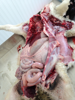
Figure 2 Exposition of the abdominal viscera during a necropsy of a lamb
Show description|Hide descriptionExposition of the abdominal viscera during a necropsy of a lamb.