
Resource 3: Working with onion cells
![]() Teacher resource for planning or adapting to use with pupils
Teacher resource for planning or adapting to use with pupils
Preparing a slide of an onion cell and measuring a cell
You will need:
Microscope
Scissors
Microscope slide
Dropper pipette
Cover slip
Clear plastic ruler
Dilute iodine solution.
Preparing the onion slide
What to do:
- Slice an onion in two, lengthwise.
- Remove one of the thick leaf-like structures from inside.
- Pull away a piece of the thin papery lining of its inner surface.
- Using scissors, cut a small square of this lining, about 5 mm x 5 mm.
- Place this square on the centre of a slide.
- Add a drop of dilute iodine solution – make sure the solution spreads below as well as above the square of onion skin. The iodine acts as a stain to make the structures in the cell easier to see.
- Carefully lower a cover slip over the onion skin. Try to avoid trapping air bubbles.
- Place the slide on the microscope stage. Examine first using the low power. Focus carefully.
- Choose an area of the slide where the cells can be clearly seen. Switch to high power and refocus.
- Look for the structures shown in the photographs in Resource 1.
Measuring the onion cell
What to do:
- Place the ruler on the microscope stage under the low power objective lens.
- Move the ruler so the edge with the scale can be focused in the centre of the field of view of the microscope, as in Diagram 1 below.
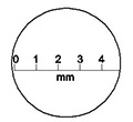
Diagram 1: The field of view of a microscope.
- Use the scale to measure the field of view of your microscope.
- The diameter of the field of view in Diagram 1 is approximately 5 mm.
- You can use the measurement of the field of view in your microscope to estimate the size of objects viewed with the same objective lens.
- The cell viewed in Diagram 2 would be about 2 mm long if viewed with the microscope with the field of view shown above.
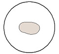
Diagram 2: Cell.
- Estimate the length and width of your onion cell using this method.
Using a microscope
The main parts of a light microscope are shown below
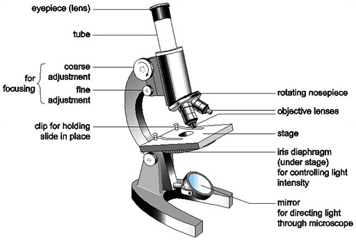
Diagram 3: Main parts of a light microscope.
Back to previous pagePrevious
Resource 2: True/false exercise on cells