
6.2.3 The pelvic outlet
The pelvic outlet is formed by the lower border of the pubic bones at the front, and the lower border of the sacrum at the back. The ischial spines point into this space on both sides. Figure 6.4 shows the dimensions of the space that the fetus must pass through as it emerges from the mother’s pelvis. As you look at Figure 6.4, imagine that you are the birth attendant who is looking up the birth canal, waiting to see the fetal head emerging.
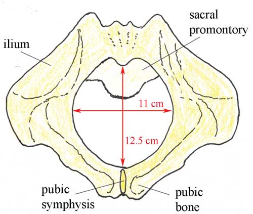
What do you notice when you compare the dimensions of the pelvic inlet (Figure 6.3) and the pelvic outlet (Figure 6.4)? Which is the narrowest?
The narrowest diameter for the fetus to pass through is the pelvic outlet, which is only 11 cm wide in the average female pelvis.
It is difficult to see from Figures 6.3 and 6.4, but the fetus has to rotate in order to get through the pelvic canal. This is because the pelvic inlet is 13 cm wide, whereas the pelvic outlet is only 11 cm wide. In order to fit through the pelvic outlet at its widest dimension (12.5 cm from top to bottom), the fetus must rotate so it ‘presents’ its head to the widest dimension of the pelvic cavity at every point as it passes through. The largest part of the fetus is the skull, so the baby’s head rotates first, and the shoulders and the rest of the body follow. You will learn all about this in the Labour and Delivery Care Module. First, we have to look more closely at the structure of the fetal skull.
6.2.2 The pelvic inlet