
Background (The structure of eukaryotic cells)
Background
In this lesson you will use a virtual light microscope to examine the structure of eukaryotic cells. Remember that living cells are divided into prokaryotes and eukaryotes. Examples of each type of cell are shown in Figure 1.

Figure 1. (a) an example of a prokaryotic cell; (b) an example of a eukaryotic cell.
|
What is the main difference you can see between the prokaryotic and eukaryotic cells shown in Figure 1? |
All animal and plant cells are eukaryotic cells. This means they consist of cytoplasm, which is bound by a cell membrane. Within the cytoplasm are other membrane-bound structures, such as the cell nucleus. Organelles perform specific functions in the cytoplasm of eukaryotic cells and include the following structures:
- cell nucleus
- mitochondrion
- vacuole
- chloroplast
- rough endoplasmic reticulum
- smooth endoplasmic reticulum
- Golgi apparatus
- lysosome
If you are unfamiliar with these terms, please refer to the Glossary section at the end of this lesson for a fuller explanation.
|
Can you think of an advantage that a cell might gain by having organelles? |
All plant and animal cells are eukaryotic cells and generalised examples of both are shown in Figure 2.
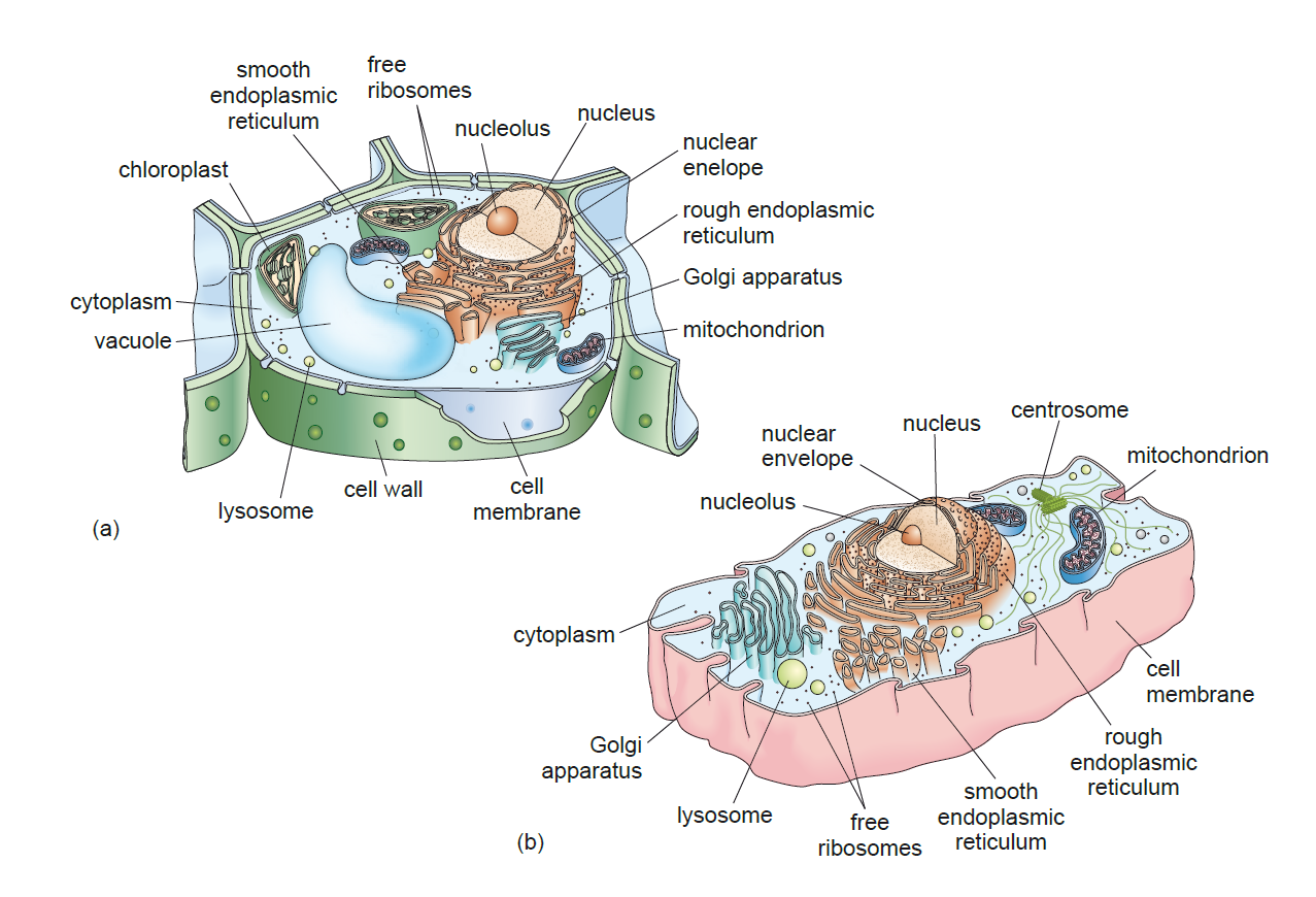
Figure 2. (a) a typical plant cell; (b) a typical animal cell.
The examples of eukaryotic cells shown in Figure 2 have been drawn in three-dimensional (3D) relief. This is so you can understand the shape and form of the cells, and structures they contain. The images you will see using the virtual microscope will be two-dimensional (2D), because they are observed in the focal plane of the microscope.
|
Activity |
|
In your notebook, redraw each of the cells shown in Figure 2 as a simple 2D image and label the structures. If you are unsure what a 2D drawing looks like, refer to the ones in Figures 3(b) and 4(b) which are 2D drawings. |
Not all organelles and cell structures are easily visible using a light microscope because the resolution is limited. Some such as mitochondria, ribosomes and the endoplasmic reticulum are best observed using an electron microscope, which uses a beam of electrons instead of light. The shorter wavelength of the electrons gives a better resolution. Figures 3 and 4 show electron micrographs of typical plant and animal cells.
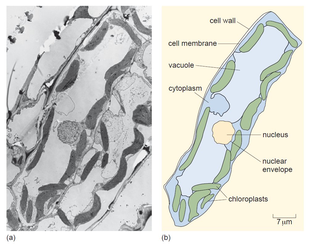
Figure 3. (a) an electron micrograph of a plant cell; (b) a 2D drawing of the plant cell shown in (a).
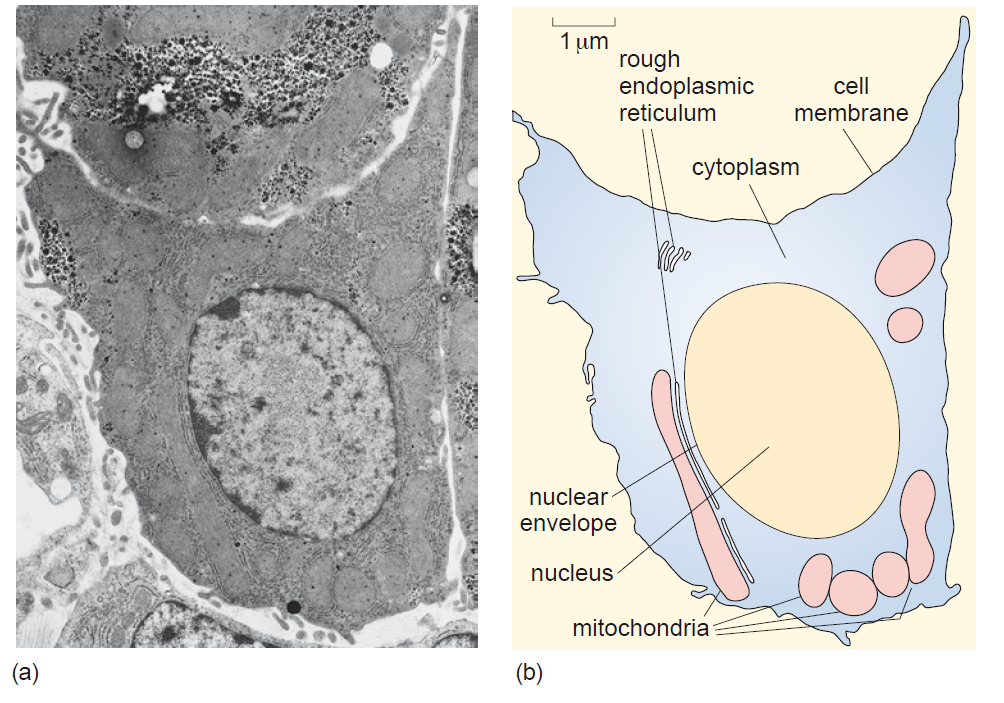
Figure 4. (a) an electron micrograph of an animal cell; (b) a 2D drawing of the animal cell shown in (a).
|
Looking at Figures 3 and 4, what two conclusions can you draw based on the images shown? (Hint: look at the scale bars) |
Now that you are familiar with the basic structure of eukaryotic cells it is time to use the virtual microscope.
Previous: Lesson objectives Next: Practical activity