4.2.1 The graph sections of the partograph
The graph sections of the partograph are where you record key features of the fetus or the mother in different areas of the chart. We will describe each feature, starting from the top of Figure 4.1 and travelling down the partograph.
- Immediately below the patient’s identification details, you record the Fetal Heart Rate initially and then every 30 minutes. The scale for fetal heart rate covers the range from 80 to 200 beats per minute.
- Below the fetal heart rate, there are two rows close together. The first of these is labelled Liquor – which is the medical term for the amniotic fluid; if the fetal membranes have ruptured, you should record the colour of the fluid initially and every 4 hours.
- The row below ‘Liquor’ is labelled Moulding; this is the extent to which the bones of the fetal skull are overlapping each other as the baby’s head is forced down the birth canal; you should assess the degree of moulding initially and every 4 hours
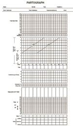
Figure 4.1 The partograph showing where to enter the patient’s identification details at the top and the graphic component below.
- Below ‘Moulding’ there is an area of the partograph labelled Cervix (cm) (Plot X) for recording cervical dilatation, i.e. the diameter of the mother’s cervix in centimetres. This area of the partograph is also where you record Descent of Head (Plot O), which is how far down the birth canal the baby’s head has progressed. You record these measurements as either X or O, initially and every 4 hours. There are two rows at the bottom of this section of the partograph to write the number of hours since you began monitoring the labour and the time on the clock.
- The next section of the partograph is for recording Contractions per 10 mins (minutes) initially and every 30 minutes.
- Below that are two rows for recording administration of Oxytocin during labour and the amount given. (You are NOT supposed to do this – it is for a doctor to decide! However, you will be trained to give oxytocin after the baby has been born if there is a risk of postpartum haemorrhage.)
- The next area is labelled Drugs given and IV fluids given to the mother.
- Near the bottom of the partograph is where you record the mother’s vital signs; the chart is labelled Pulse and BP (blood pressure) with a possible range from 60 to 180. Below that you record the mother’s Temp °C (temperature).
- At the very bottom you record the characteristics of the mother’s Urine: protein, acetone, volume. You learned how to use urine dipsticks to test for the presence of a protein (albumin) during antenatal care.
You learned about giving IV (intravenous) fluid therapy to women who are haemorrhaging in Study Session 22 of the Antenatal Care Module.
What can you tell from the colour of the amniotic fluid?
If it has fresh bright red blood in it, this is a warning sign that the mother may be haemorrhaging internally; if it has dark green meconium (the baby’s first stool) in it, this is a sign of fetal distress.
Back to previous pagePrevious
4.2 Finding your way around of the partograph