
3.1.2 Olpidiopsis disease
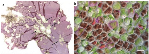
The disease shown in Figure 10 has been reported in China, Korea and Japan. Initially thought to be a chytrid, Olpidiopsis is actually an obligate endoparasitic oomycete, infecting many different species of Pyropia. Symptoms (seen in Figure 10) are characterised by distinct bleached portions on blades at the initial stages and the development of greenish lesions upon spreading of the disease. The infection process starts when encysted zoospores of Olpidiopsis attach to the surface of Pyropia and produce thin germ tubes that penetrate the cell walls of the host. After entering the host cell, Olpidiopsis forms spherical multinucleate thalli, which develop into fully grown zoosporangia within the next two days. Zoospores escape through a discharge tube, which forms at the final developmental stage of the zoosporangium. Again, decay of tissue in infected areas quickly promotes the death of the whole plant.
3.1.1 Pyropia Red Rot disease