
3.4.3 Scenedesmus dimorphus
The production of biofuels using algae is an attractive technology that could mitigate the ongoing depletion of fossil reserves and their impact onimpacts/influence on climate change. Open pond mass cultivation of the green alga Scenedesmus dimorphus is considered as one of the most promising strategy for cost effective production of algae. Again, with the recent implementation of those culture strategies, multiple reports of parasitic attacks that can destroy mass cultures in a matter of days have been reported raising concern for this developing sector. Amoeboaphelidium protococcarum belong to the Cryptomycota clade and has been recently described in a study by Letcher et al, 2014 [Tip: hold Ctrl and click a link to open it in a new tab. (Hide tip)] . Its life cycle is illustrated in Figure 16.
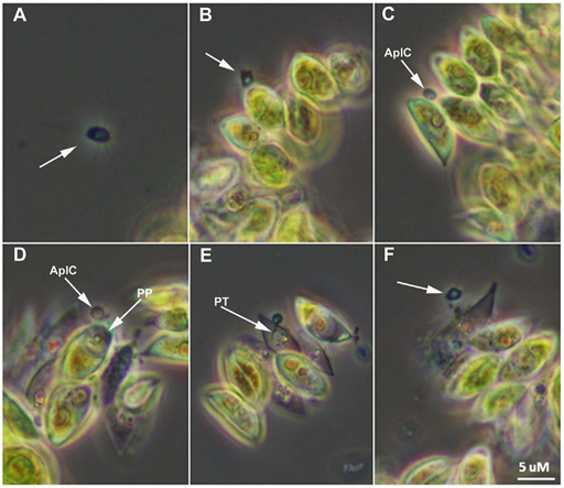
In the figure, A. Motile aplanospore (arrow) is proximal to host. B. Aplanospore (arrow) encounters Scenedesmus dimorphus cells and attaches. C.
Aplanospore has completed attachment and has formed an epibiotic cyst (AplC). D.
Parasite protoplasm (PP) has been injected into the host cell and clearly visible within the cell. E. Clear view of the parasite penetration tube (PT); progeny begin to mature within parasite sporangium. F. Dehiscence of aplanospore cyst occurs and progeny (arrow) are released from the infected cell.
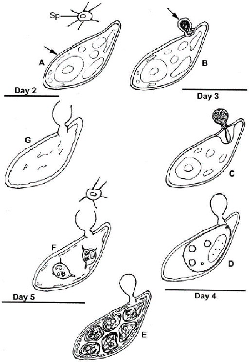
At Day 2: A. Abundant healthy algal cells (arrow) and abundant amoeboid, filose pseudopodiate aplanospores (Sp) are evident. B. A minority of algal cells indicate infection by the presence of a cyst (arrow) attached via an appressorium to the algal cell wall.
Day 3: C. The walled, stalked aplanospore cyst has enlarged, a penetration tube penetrates the host cell wall, and a host reaction at the site of infection is evident. D. In some algal cells a membrane-bound, developing parasite protoplasm occupies a portion of the interior of the host cell, and a host/parasite interface is evident; the empty remnant of the aplanospore cyst persists.
Day 4: E. Cleaved aplanospores within the host cell are surrounded by fused and fragmented plasma membrane; the empty remnant of the aplanospore cyst persists.
Day 5: F. A minority of host cells retain one or a few unreleased aplanospores, a sub-apical portion of the empty remnant of the aplanospore cyst has dissolved, and abundant aplanospores occur outside host cells. G. Most infected host cells are empty, but retain the remnant of the aplanospore cyst.
Further reading
If you want to learn more about the biology of Aphelids and the ongoing work concerning their classification, you could read the following articles:
3.4.2 Microalgae