
Science, Maths & Technology
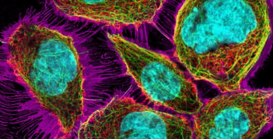
Exploring cells with digital fluorescence microscopy
...system. This allows for the capture of a series of optical sections – thin, focused slices – at different depths within a specimen. These images form a z-stack (Figure 15b), which computer software can reconstruct into a highly detailed three-dimensional (3D) image of the cell or tissue (Figure 15c). This is particularly useful for studying complex structures in thick...


