Skip to content
Skip to main content

About this free course

Download this course

Share this free course

Understanding science: what we cannot know

Start this free course now. Just create an account and sign in. Enrol and complete the course for a free statement of participation or digital badge if available.

This is a collage of four images of the human brain, all showing that it has a walnut-like appearance of ridges and grooves across its surface. Figure 1 (a) is a diagram of a human brain located inside a head, showing the large cerebrum occupying most of the skull, with the smaller cerebellum underneath the back of the cerebrum, and the brain stem emanating from under the cerebrum, just in front of the cerebellum. The cerebrum is responsible for much of the complex, higher order processing that takes place in the brain, while the cerebellum is responsible for controlling balance and movement. The brain stem is responsible for controlling involuntary processes such as breathing and heart rate. Figure 1 (b) is a colour photograph of the human brain, viewed from above. The two semi-oval (in the photograph) cerebral hemispheres are visible, right and left, with the longitudinal fissure between them. The surface of the brain (grey in the photograph) shows extensive gyri and sulci (ridges and grooves). Figure 1 (c) contains two images. The left image shows a sagittal view of a model of the human brain with the front of the brain towards the left and the back of the brain towards the right. The corpus callosum is highlighted as an elongated c-shaped structure, rotated 90 degrees clockwise and it sits just below the cerebral cortex. The anterior is almost level with the pituitary gland and the posterior is level with the back of the brain stem. It therefore occupies the middle region of the brain, from front to back. The right image in Figure 1 (c) is a colour photograph of a post-mortem human brain sample, viewed from above, with the top layer of cortex removed to show the thick band of the corpus callosum with numerous fibres projecting laterally across the midline of the brain from one hemisphere to another.
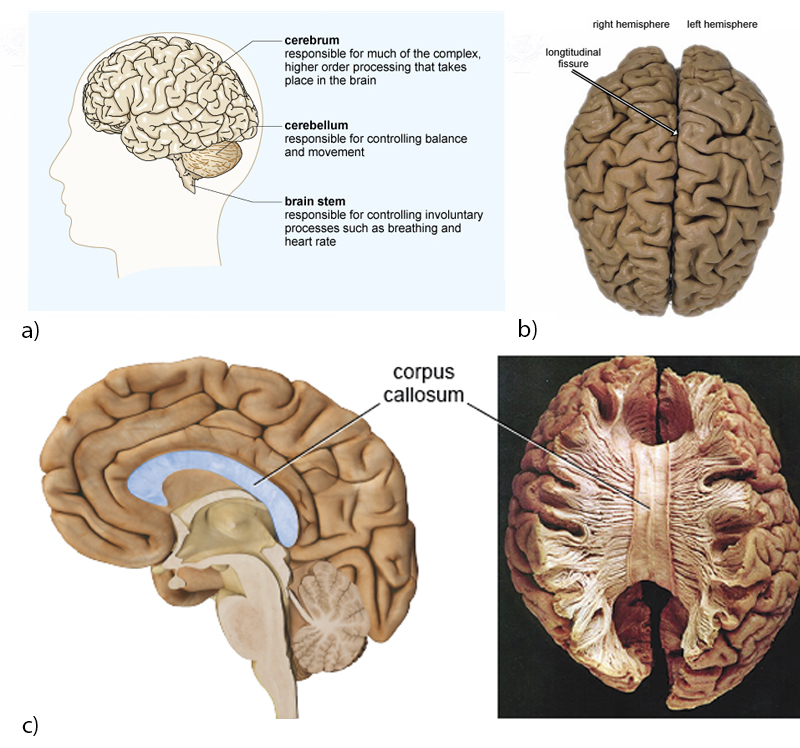
Figure 1 (a) the human brain viewed from the side and (b) in a photograph from above. (c) Left: a cut-away view through the middle of the brain. Right: A post-mortem human brain sample with top layer removed.

 2.1 Anatomy of the brain