Skip to content
Skip to main content

About this free course

Download this course

Share this free course

Understanding science: what we cannot know

Start this free course now. Just create an account and sign in. Enrol and complete the course for a free statement of participation or digital badge if available.

2.2 Neurons and neurotransmitters

Meet ‘neurons’. These are what would colloquially be referred to as ‘brain cells’, although the brain is made up of many other types of cells as well. This short video introduces the cells’ structure and function.

Download this video clip.Video player: Video 4
Copy this transcript to the clipboard
Print this transcript
Show transcript|Hide transcript
Video 4 Neurons
Interactive feature not available in single page view (see it in standard view).

As you’ve just seen, neurons are special cells in the brain which transmit electrical pulses. This is how the different parts of the brain ‘talk’ to each other, and indeed to the rest of the body via other neurons in the ‘nervous system’. You can think of this as being like the body’s electrical wiring.

Neurons in the brain form a communication network, as illustrated in this short video clip.

Download this video clip.Video player: Video 5
Copy this transcript to the clipboard
Print this transcript
Show transcript|Hide transcript
Video 5 Neuron networks
Interactive feature not available in single page view (see it in standard view).

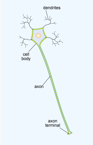
The diagram in Figure 2 illustrates a typical neuron. The microscopic cell body contains important cellular components, such as the nucleus that houses genetic material. Typically, the branching dendrites receive incoming messages from neighbouring neurons, which are then integrated together. If the neuron ‘fires’, the axon carries the signal as an electrical pulse to the axon terminals. The length of an axon varies enormously, from a fraction of a millimetre to a metre or more. The longest axons in humans belong to neurons in the sciatic nerve, which connects the base of the spine to the toes.

This diagram shows a schematic representation of a neuron, with its key features labelled. Towards the top is the small cell body which is surrounded by a membrane and which contains a nucleus in its centre. Extending from around the cell body are several small tree-like structures with a simple branched form, labelled as dendrites. Also extending down from the cell body is a long thin axon. At the end of the axon is the axon terminal which is a slightly widened feature.
Figure 2 The main structural features of a typical neuron

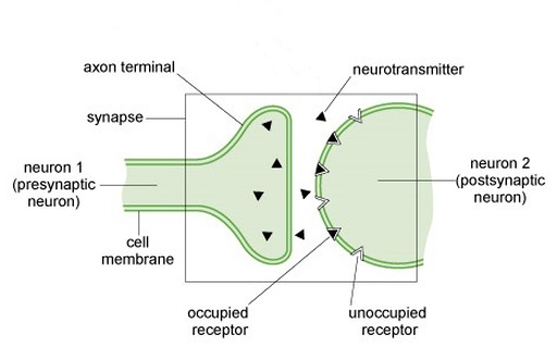
The connections between neurons are called ‘synapses’. In most cases, a signal is transmitted across the small gaps between neurons by chemicals called neurotransmitters, rather than by electrical pulses. Some neurotransmitters act to ‘excite’ neurons while others have an inhibitory effect, but if the overall chemical signal is strong enough then adjacent neurons will ‘fire’. There are many different neurotransmitters, but you may be familiar with a few names already, like adrenaline, endorphin, dopamine and histamine.

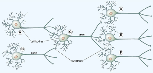
The diagram in Figure 3 shows one neuron releasing chemical neurotransmitters, triggered by the electrical pulses travelling along the axon. The surface membrane of the next neuron contains specific receptors for these neurotransmitters, acting somewhat like a lock-and-key mechanism. These chemical messages can be passed to the branching dendrites of more than one neuron, like in the simple network shown in Figure 4.

This diagram shows some detail of the molecular events at the synapse. The bulb-shaped ends of two neurons are shown in close proximity to each other. The region encompassing them together with the gap between them is labelled the synapse. The axon terminal of the neuron on the left is depicted as containing small triangular-shaped neurotransmitters, some of which are crossing the gap towards the neuron on the right. The bulbed end of the neuron on the right is depicted with complementary triangular shaped wedges cut out. Some of these are filled by the neurotransmitters. These are labelled as unoccupied receptors and occupied receptors, respectively.
Figure 3 A schematic representation of a synapse
This diagram shows six neurons connected together in a network (labelled A–F). They are positioned in a horizontal orientation with the dendrites and cell body to the left and the axons extending towards the right. Each axon branches into three, and theses axon terminals are connected to the dendrites of another neuron or neurons positioned to the right of it. The point where the axons and dendrites are connected are labelled synapses. From left to right, two neurons are connected to one neuron, which is in turn connected to three neurons. Neuron A lies on the top left of the diagram, with its dendrites on the left and the axon extending horizontally towards the right. Neuron B lies immediately below this in the same orientation. Neuron C is to the right of neurons A and B, and again is lying in a horizontal orientation with its cell body on the left and axon extending towards the right. The axon terminals of neurons A and B form synapses with the dendrites of neuron C. To the right of neuron C are three neurons (D, E and F) lying horizontally and aligned one above the other. The axon terminals of neuron C forms synapses with the dendrites of neurons D, E and F.
Figure 4 A simple network of neurons, in which information is passed from left to right

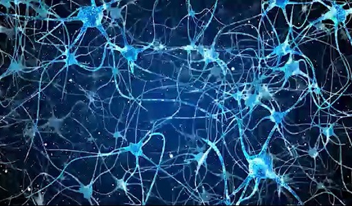
Some neurons can ‘pass on the message’ to hundreds or even thousands of other neurons in this way. With around 100 billion neurons in the brain – similar to the number of stars in an entire galaxy – this makes many trillions of connections in total! And it isn’t just the number of connections which is astounding, but the number of messages which are being sent – a neuron can fire off hundreds of messages each second. Video 6 visually demonstrates a few of this section’s concepts.

Download this video clip.Video player: Video 6
Copy this transcript to the clipboard
Print this transcript
Show transcript|Hide transcript
Video 6 How neurons communicate
Interactive feature not available in single page view (see it in standard view).