Skip to content
Skip to main content

About this free course

Download this course

Share this free course

Understanding science: what we cannot know

Start this free course now. Just create an account and sign in. Enrol and complete the course for a free statement of participation or digital badge if available.

3.1 Microscopy and silver nitrate staining

Brain tissue can be treated with chemicals to stain some of the individual neurons, allowing their finely detailed structure to be seen with a microscope. This technique was discovered in the 1870s by Camillo Golgi, and later used and improved by Santiago Ramón y Cajal. It enabled major advancements in neuroscience, particularly in identifying neurons as the key to brain function. The biologists were awarded the Nobel Prize ‘in recognition of their work on the structure of the nervous system’ in 1906. Figure 7 shows ‘Golgi stained’ brain tissue viewed under a microscope, with Figure 7a revealing the structure of individual neurons, and 7b showing the patterns of neurons in a section of a mouse brain.

This is a composite of two images of brain tissue taken through a microscope in which neurons are clearly visible as black structures against a lighter or white background. In image (a), three clearly distinguishable neurons are seen with distinctive pyramidal-shaped cell bodies, branching dendrites and a single axon projecting away from the cell body. Further cells are visible in the background but these can’t be clearly identified. In image (b), ‘forests’ of many neurons are seen in distinct roughly horizontal curving layers. The layers can be distinguished by differences in the density and morphology of the neurons. There are also distinct gaps between some layers where almost no neurons are seen.
Figure 7 ‘Golgi stained’ brain tissue viewed under a microscope

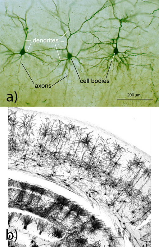
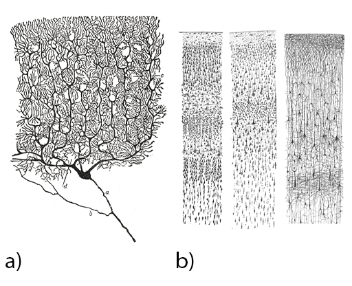
Figure 8a shows a single ‘Purkinje’ neuron, one of the largest cells in the brain, with an elaborate structure of branched dendrites. Figure 8b shows interconnected neurons in the cortex, appearing in layers.

This is a composite of two highly detailed black and white drawings of neurons. Image (a) shows a single large and very elaborate neuron. It has a tree-like structure, with a blob-shaped cell body, an axon projecting downwards, and numerous highly branched dendrites forming the tree ‘canopy’. Image (b) consists of three drawings showing ‘forests’ of neurons appearing in horizontal layers. The layers can be distinguished by differences in the density of the neurons. Within each layer there are cell bodies of neurons and for some layers, the axons can be seen running vertically from one layer to the next. The drawing on the right particularly gives a sense of a dense connectivity between neurons.
Figure 8 Neurons in ‘Golgi stained’ brain tissue (drawings by Santiago Ramón y Cajal)

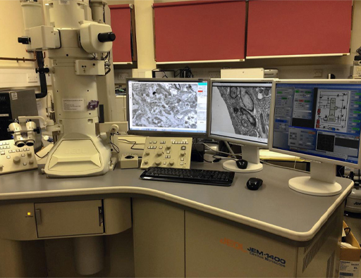
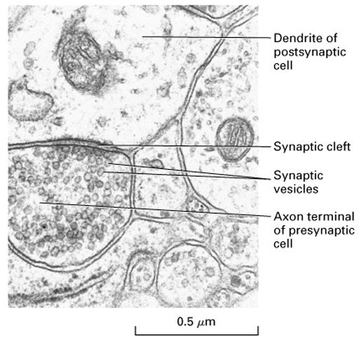
The electron microscope was invented in the 1930s and developed over subsequent decades. Rather than light, a beam of subatomic electrons is used to image objects, allowing for much greater magnification, and visualisation on the nanometre scale. This invention was a significant step for neuroscience. At the end of the 1950s, individual synapses were imaged, cementing the neuron theory of brain function.

This is a photograph of a scientific laboratory. On a workbench on the left is the electron microscope, which looks like a large vertical metal tube-shaped structure with various knobs and attachments. To the right of this are three computer screens showing different magnified outputs from the electron microscope and control screens.
Figure 9 Electron microscope
This is an image taken through an electron microscope showing the internal features in the region of a synapse. The image is highly magnified and contains a number of bubble-like structures of different sizes. One bubble to the lower left is the axon terminal of the presynaptic neuron. The scale indicates that it has a diameter of about half a micrometre. It is filled with numerous small round structures which are the synaptic vesicles containing neurotransmitters. Above and in close contact with this is part of a dendrite of postsynaptic neuron. It is filled with some darker blobs of varying sizes which are not labelled. Separating the pre- and postsynaptic neurons is a gap called the synaptic cleft. There are some other similar structures in the image which aren’t identified.
Figure 10 An electron micrograph showing synapses. The synaptic cleft is the gap between the neurons, and the synaptic vesicles in the presynaptic neurons contain neurotransmitters.