

2 The anatomy of a virus
Viruses come in a variety of different shapes and sizes. The simplest consist of a protein shell called the nucleocapsid, which contains the viral genetic material (nucleic acid). These are called non-enveloped viruses, to distinguish them from the more complex enveloped viruses, as shown in Figure 2.

Enveloped viruses also have a nucleocapsid containing the viral genome. The nucleocapsid is contained within a viral envelope, a composite structure which includes a phospholipid bilayer, derived from the plasma membrane of the host cells which produced the virus. Inside the membrane, viral matrix proteins connect the nucleocapsid to the envelope. The matrix is also important in organising the assembly of new virus particles. The envelope also contains viral proteins and some residual host proteins from the infected cell.
The critical thing to notice is that some proteins are on the outside of the virus whereas others, such as the nucleocapsid and matrix proteins, are on the inside. This is important because it affects what parts of the virus can be recognised by different elements of the immune system.
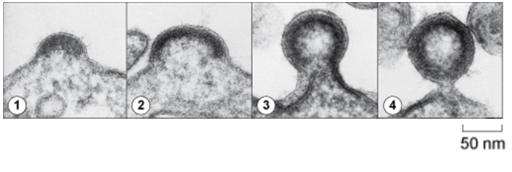
As you can see in Figure 3, enveloped viruses are released from infected cells by budding from the plasma membrane. Examples of enveloped viruses are influenza-A, HIV and SARS-CoV2. The transmission electron micrographs show different stages in the assembly (1, 2) budding (3) and release (4) of HIV from the surface of an infected cell. This process illustrates step 4 in Video 2.
In addition to the components described above, most viruses contain a number of enzymes and auxiliary proteins which are required to initiate infection of the cell and replication of the virus.