

1.3 Antigen processing and presentation
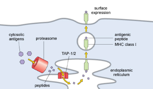
As previously noted, a cell samples its internal proteins and presents them on MHC class I molecules. The way it does this is called ‘antigen processing’ and is illustrated in Figure 3. Cells have an organelle called a proteasome, which breaks down cytosolic proteins into peptides that are transported into the endoplasmic reticulum by a transporter (TAP1/2), where they can associate with MHC class I molecules.
ITQ _unit3.2.3
-
What determines whether a peptide will be able to bind to an MHC molecule?
-
The amino acid residues lining the antigen-binding cleft on the MHC molecule interact with the amino acid residues in the peptide, so binding depends on both the variant of the MHC molecule and the sequence of the peptide.
The antigenic peptides are trimmed to size by enzymes in the endoplasmic reticulum before the MHC molecule is transported to the plasma membrane in order to present the antigen.