
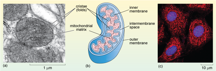
Figure 22 (a) Electron micrograph of mitochondria from a rat liver cell. The double membrane and cristae are clearly visible. (b) Schematic diagram of a mitochondrion. (c) Fluorescent light micrograph of cultured human cells. The nuclei are labelled blue and the mitochondria red. The image shows the irregular shapes and network-like arrangement of the mitochondria in these cells.