Skip to content
Skip to main content

About this free course

Download this course

Share this free course

Understanding antibiotic resistance

Start this free course now. Just create an account and sign in. Enrol and complete the course for a free statement of participation or digital badge if available.

4.1 Gram-positive and Gram-negative bacteria

Bacteria are divided into two groups based on how the cell wall appears when they are stained using Gram straining. This procedure allows the composition of the wall to be visualised.

In Gram-positive bacteria, the cell wall has a thick peptidoglycan layer which is relatively porous, allowing substances to pass through it quite easily.

In Gram-negative bacteria, this peptidoglycan layer is greatly reduced and is further protected by a second, outer membrane (Figure 10).

Arrangement of the cell wall in (a) Gram-positive and (b) Gram-negative bacteria.
Figure 10 Arrangement of the cell wall in (a) Gram-positive and (b) Gram-negative bacteria.

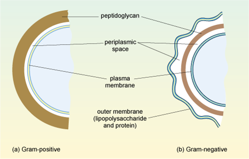
This second, outer membrane of Gram-negative bacteria is an effective barrier, regulating the passage of large molecules such as antibiotics into the cell. In contrast, the thick, porous peptidoglycan layer in the cell wall of Gram-positive bacteria gives greater access to antibiotics, allowing them to more easily penetrate the cell and/or interact with the peptidoglycan itself.

You will learn more about the strategies antibiotics use to cross the cell wall in Week 3.