1.2.1 The circulation of blood
The heart is a large four-chambered muscular bag on the left side of the chest. In order to appreciate how the heart works, remind yourself of the primary function of the cardiovascular system: to deliver oxygen and nutrients and to remove carbon dioxide and other waste products. When you breathe in, the lungs are filled with air, of which about 21% is oxygen. To collect this oxygen, the blood has to be pumped through the lungs by the heart.
Oxygenated blood (blood rich in oxygen) from the lungs, which is bright red because oxygen has bound to the haemoglobin, returns to the heart and is then pumped around the body to supply the tissues. Blood returning from the body to the heart is rich in waste products such as carbon dioxide and is short of oxygen. This oxygen-depleted blood (dark red in colour) is termed deoxygenated blood and is pumped through the lungs again to release carbon dioxide and, of course, to collect more oxygen.
The design of the heart and associated blood vessels ensures that blood going to the lungs is kept separate from that going around the body. The heart prevents the mixing of oxygenated blood with deoxygenated blood by using two separate but parallel circuits of blood vessels: the pulmonary circulation and the systemic circulation.
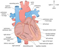
Because of its four-chamber design, the heart can serve both circuits at once, using its two pumps to simultaneously push blood from one circuit through one half of its structure and blood from the other circuit through its other half (see Figure 1.3).
The muscular part of a heart is called the myocardium (‘myo-’ means ‘muscle’ and ‘-cardium’ means ‘of the heart’). The heart muscles are very special because they keep beating (contracting and relaxing) spontaneously throughout our whole lives without any conscious decision from us to make them beat.
As you can see from Figure 1.3, the heart is shown in cross-section, illustrating the position of the atria, ventricles and major veins and arteries. The left and right sides of the heart are separated by a muscular wall (called the septum), and each side is divided into a small chamber, the atrium (plural, atria), and a larger chamber, the ventricle (plural, ventricles). The atria are connected to the ventricles via a valve that ensures a one-way flow of blood. Deoxygenated blood returns from the body through two main ‘great’ veins, the inferior and superior vena cava (superior means ‘at the top’ and inferior means ‘at the bottom’ as you can see from their positions in Figure 1.3).
The atrium is a thin-walled chamber that expands with little resistance as the blood enters. Blood from the right atrium flows down into the right ventricle, through the tricuspid valve. You can imagine the valve operating in a manner similar to a swing door that only opens in one direction. When blood enters the right atrium, the valve opens and blood flows into the right ventricle. When the ventricles contract, the back pressure of the blood forces the valve to close to prevent any backflow of blood into the atria.
1.2 Anatomy and physiology of the heart