
This content is associated with The Open University's Health Science courses and qualifications.
What is FMRI?
![Brain image produced by FMRI [Image: FMRIB Centre]](https://www.open.edu/openlearn/pluginfile.php/3259025/tool_ocwmanage/articletext/0/fp_image.jpg) Functional magnetic resonance imaging, or FMRI, works by detecting the changes in blood oxygenation and flow that occur in response to neural activity – when a brain area is more active it consumes more oxygen and to meet this increased demand blood flow increases to the active area. FMRI can be used to produce activation maps showing which parts of the brain are involved in a particular mental process.
Functional magnetic resonance imaging, or FMRI, works by detecting the changes in blood oxygenation and flow that occur in response to neural activity – when a brain area is more active it consumes more oxygen and to meet this increased demand blood flow increases to the active area. FMRI can be used to produce activation maps showing which parts of the brain are involved in a particular mental process.
Background
The development of FMRI in the 1990s, generally credited to Seiji Ogawa and Ken Kwong, is the latest in long line of innovations, including positron emission tomography (PET) and near infrared spectroscopy (NIRS), which use blood flow and oxygen metabolism to infer brain activity. As a brain imaging technique FMRI has several significant advantages:
- It is non-invasive and doesn’t involve radiation, making it safe for the subject.
- It has excellent spatial and good temporal resolution.
- It is easy for the experimenter to use.
The attractions of FMRI have made it a popular tool for imaging normal brain function – especially for psychologists. Over the last decade it has provided new insight to the investigation of how memories are formed, language, pain, learning and emotion to name but a few areas of research. FMRI is also being applied in clinical and commercial settings.
![Brain showing areas responsible for sense [Image: FMRIB]](https://www.open.edu/openlearn/pluginfile.php/3259025/tool_ocwmanage/articletext/0/brainschematic.jpg)
Sensory regions of the brain - Courtesy of Peter Hobden, FMRIB
How does MRI work?
FMRI is a special type of magnetic scan.
The cylindrical tube of an MRI scanner houses a very powerful electro-magnet. A typical research scanner (such as the FMRIB Centre scanner) has a field strength of 3 teslas (T), about 50,000 times greater than the Earth’s field.
The magnetic field inside the scanner affects the magnetic nuclei of atoms. Normally atomic nuclei are randomly oriented but under the influence of a magnetic field the nuclei become aligned with the direction of the field. The stronger the field the greater the degree of alignment.
When pointing in the same direction, the tiny magnetic signals from individual nuclei add up coherently resulting in a signal that is large enough to measure. In FMRI it is the magnetic signal from hydrogen nuclei in water (H2O) that is detected.
The key to MRI is that the signal from hydrogen nuclei varies in strength depending on the surroundings. This provides a means of discriminating between grey matter, white matter and cerebral spinal fluid in structural images of the brain.
What does FMRI measure?
Oxygen is delivered to neurons by haemoglobin in capillary red blood cells. When neuronal activity increases there is an increased demand for oxygen and the local response is an increase in blood flow to regions of increased neural activity.
Haemoglobin is diamagnetic when oxygenated but paramagnetic when deoxygenated. This difference in magnetic properties leads to small differences in the MR signal of blood depending on the degree of oxygenation. Since blood oxygenation varies according to the levels of neural activity these differences can be used to detect brain activity. This form of MRI is known as blood oxygenation level dependent (BOLD) imaging.
![The BOLD effect [Image: FMRIB]](https://www.open.edu/openlearn/pluginfile.php/3259025/tool_ocwmanage/articletext/0/boldeffect.jpg)
Diagram of the BOLD effect - Courtesy of Stuart Clare, FMRIB
One point to note is the direction of oxygenation change with increased activity. You might expect blood oxygenation to decrease with activation, but the reality is a little more complex.
There is a momentary decrease in blood oxygenation immediately after neural activity increases, known as the “initial dip” in the haemodynamic response. This is followed by a period where the blood flow increases, not just to a level where oxygen demand is met, but overcompensating for the increased demand.
This means the blood oxygenation actually increases following neural activation. The blood flow peaks after around 6 seconds and then falls back to baseline, often accompanied by a "post-stimulus undershoot".
Activation maps
![Audio visual FMRI - single slice [Image: FMRIB]](https://www.open.edu/openlearn/pluginfile.php/3259025/tool_ocwmanage/articletext/0/activationmap.jpg) This image [left, courtesy Steve Smith, FMRIB] is the result of the simplest kind of FMRI experiment. While lying in the MRI scanner the subject watched a screen which alternated between showing a visual stimulus and being dark every 30 second. Meanwhile the MRI scanner tracked the signal throughout the brain.
This image [left, courtesy Steve Smith, FMRIB] is the result of the simplest kind of FMRI experiment. While lying in the MRI scanner the subject watched a screen which alternated between showing a visual stimulus and being dark every 30 second. Meanwhile the MRI scanner tracked the signal throughout the brain.
In brain areas responding to the visual stimulus you would expect the signal to go up and down as the stimulus is turned on and off, albeit blurred slightly by the delay in the blood flow response. The 'activity' in a voxel (the smallest unit in a three-dimensional image) is defined as how closely the time-course of the signal from that voxel matches the expected time-course.
Voxels whose signal corresponds tightly are given a high activation score, voxels showing no correlation have a low score and voxels showing the opposite (deactivation) are given a negative score. These can then be translated into activation maps.
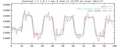
Time course graph courtesy of Steve Smith, FMRIB
Images from FMRI experiments are often presented in colour to make it easier to visualise results.
Clinical and commercial use
FMRI now has a small but growing role in clinical neuroimaging. It is used in pre-surgical planning to localise brain function.
There is also potential for clinical FMRI in applications including presymptomatic diagnosis, drug development, individualisation of therapies and understanding functional brain disorders. Early studies also suggest that FMRI has the potential to be used as bio-feedback for conditions such as chronic pain.
There have been several early ventures to capitalise on FMRI. Two companies have been set up in North America offering lie detection services using FMRI. There are also several neuromarketing companies, using FMRI to gain insights into consumer thought and behaviour.
How FMRI Works is adapted, with permission, from an article produced by the University of Oxford FMRIB Centre. You can read the full introduction to FMRI on their website.
Study a free health course
-
Exploring health: is your lifestyle really to blame?
Learn more to access more details of Exploring health: is your lifestyle really to blame?This free course, Exploring health: is your lifestyle really to blame?, explores the extent to which a person's 'lifestyle' impacts on their health and wellbeing. It also examines how non-lifestyle related factors – in particular social, economic, cultural and political dimensions – influence a person's health.

-
Population ageing: a global health crisis?
Learn more to access more details of Population ageing: a global health crisis?This free course, Population ageing: a global health crisis?, focuses on two major issues of our time – ageing societies and global health. It provides you with an introduction to ageing societies and their implications for global health – implications which are only just beginning to be fully understood. The course will help you to deepen your ...

Discover more content like this
-
Beachbrains
Read now to access more details of BeachbrainsEver tried to picture how many neurons we have inside our brain but can't envisage it? Use this busy beach analogy to help you understand the brain:

-
The delivery service to fix your brain
Read now to access more details of The delivery service to fix your brainHow can we make sure drugs get to where they are needed in the body? Open University PhD student Conor McQuaid explains one way in which scientists can target the delivery of drugs.

Rate and Review
Rate this article
Review this article
Log into OpenLearn to leave reviews and join in the conversation.
Article reviews