Skip to content
Skip to main content

About this free course

Download this course

Share this free course

Understanding science: what we cannot know

Start this free course now. Just create an account and sign in. Enrol and complete the course for a free statement of participation or digital badge if available.

Imaging

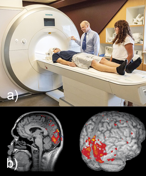
New imaging techniques give us more detailed ‘snapshots’ of what’s going on inside the brain. These don’t measure the electrical activity of neurons directly, but measure other things associated with brain activity. Active regions in the brain require more oxygen, which is supplied by an increased blood flow. This can be measured in various ways, but the most common technique uses the fact that the increased oxygenation changes the magnetic properties of the blood. This is detected non-invasively by an fMRI (functional Magnetic Resonance Imaging) scanner. Figure 12 shows such a scanner, and a scan showing regions in the brain that become active in response to a visual stimulus.

This is a composite of two images, showing an fMRI scanner and some examples of fMRI scans. Image (a) is a photograph in which a patient is about to enter a medical scanner. The patient is lying on a medical bench which is part of a large white machine with a tunnel at his head. His head is held in place by a frame. Two medical researchers are standing next to him. Image (b) shows two images taken by an fMRI scan. Both are greyscale images of brain tissue but with some regions of red and orange superimposed on top, indicating increased brain activity in these regions. The image on the left is a side view of a human brain inside a head, looking to the left. Structures in the centre of the brain are visible, including the convoluted surface of the cerebrum and cerebellum, the brain stem and corpus callosum. The skull surrounding the brain, and bones and soft tissue of the face can also be identified. The image on the right shows an external view of brain taken from the rear. Only the external features of the brain can be viewed in this image, in particular, the ridges and grooves of the cerebral cortex. In both images, there is a region at the back of the cerebrum in the occipital lobe which appears red and orange.
Figure 12 (a) fMRI scanner, (b) fMRI scan

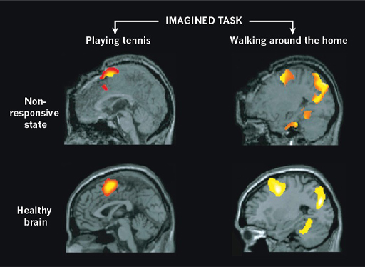
Researchers can observe active regions in the brain associated with carrying out tasks or having different experiences, including sensing pain, using language, storing memories, feeling particular emotions, and so on. These same regions are even activated when a subject is asked to imagine doing something. In Figure 13, brain activity in an area associated with movement is seen when the subject imagines playing tennis, and activity in areas associated with movement and memory is seen when the subject imagines walking through their own home. Similar brain activity is also seen when some apparently non-responsive patients are asked to imagine these tasks.

Four images taken by an fMRI scan are shown. These are greyscale images of brain tissue with some regions of red, orange and yellow superimposed on top showing regions of brain activity. Each is a side view of a human brain inside a head, looking to the left. The two images on the left are from different patients imagining playing tennis, while the two images on the right are from patients imagining walking around the home. In each case one patient has a healthy brain and the other patient is in a non-responsive state. For each imagined task, the regions of brain activity are very similar in both patients.
Figure 13 fMRI scans showing brain activity while tasks are imagined

Each individual ‘voxel’ (like a 3-dimensional pixel) of current fMRI images still covers a broad region of the brain, encompassing many thousands of neurons. But this resolution is improving over time, allowing more and more detailed pictures of brain processes.