3.3 Into the future
A triumph of modern biological science has been mapping the human genome – the complete genetic code that is contained in our DNA, with over 3 billion units of information. The Human Genome Project was completed in 2003, after 13 years of unsurpassed international collaboration. Another related quest is the Human Connectome Project, which is ongoing at time of writing. It aims to construct a map of all the neural connections in the brain, termed a ‘connectome’. This could be thought of as a ‘wiring diagram’ for the brain.
Limited mapping of neural circuits is currently possible, using the microscopy techniques on brain tissue seen in Section 3.1. But even with recent refinements in methodology, mapping the whole brain and its 86 billion neurons would be an impossibly vast task. It would take over a year to acquire the data for just 1 cubic millimetre of brain tissue, and the raw data for the whole brain would require computing storage of about 175 exabytes – that’s 175 billion gigabytes!
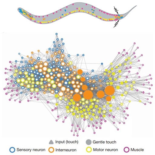
What has been possible, though, is the mapping of the entire connectome of a much simpler creature: a transparent worm just one millimetre long.
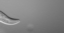
This worm doesn’t have a brain – it’s controlled by a nervous system containing just a few hundred neurons. Nevertheless, it took researchers over a decade of work to produce this ‘circuit diagram’.
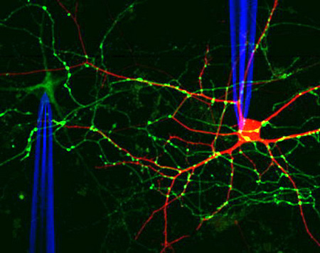
The connective architecture of animal brains can be studied on larger scales with a different approach. This involves injecting ‘tracer molecules’ that are then transported along the axons of neurons. Typically, these compounds are fluorescent, rendering the pathways visible in a conventional microscope.
Figure 17 shows neuron (in red) made visible by the introduction of a dye using a microelectrode. A second neuron is traced using a fluorescent protein – the green dots show the locations of individual synapses.
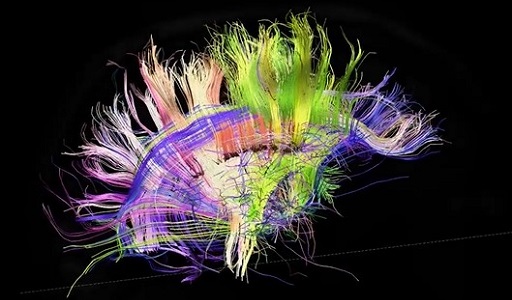
A further technique for studying human brains is to use non-invasive MRI (magnetic resonance imaging) to track how water diffuses through the brain, and so trace the main neural pathways (consisting of hundreds of thousands of axons).
It turns out that the connective structure of the brain is remarkably ordered, as shown in this video.