2.6 Communication: functions of the nervous system
The function of the nervous system, and the cells that comprise it, is to communicate signals that originate both within the body and in the external environment.
As you read earlier in the course, the nervous system is divided into two main parts.
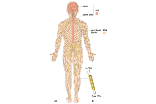
- the central nervous system (CNS) includes the brain, spinal cord and retina of the eye
- the peripheral nervous system (PNS) is the network of nerves outside the CNS that runs through virtually all other tissues.
Both parts are illustrated in Figure 12.

There are a greater abundance of nerve cells in some parts of the body than others. For example, there is a dense plexus of nerves associated with the gut, which is know as the enteric nervous system. This network of cells controls peristalsis – the coordinated contraction of the muscles along the gut wall that moves matter through the digestive system.
The roles and structures of neurons
Around half of the cells that comprise the nervous system are called neurons.
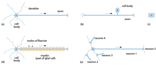
Neurons typically consist of a cell body, which contains the nucleus of the cell. Dendrites extend from the cell body, and this is where information (signals) are received. Each cell also has a single axon, which relays signals onwards to other neurons or to other cells such as muscle. A number of possible arrangements of neural structures are depicted below in Figure 13.

The roles and structures of other cell types
The other cells that comprise the nervous system perform supporting functions for the neurons, and are collectively known as glia. Examples of glia are astrocytes, oligodendrocytes, Schwann cells and microglia, and these are described in more detail below.
Astrocytes control many metabolic functions within the nervous system and they signal between neurons and the vascular system to control blood flow.
Many nerves have a sheath of insulating material (myelin) around their axons, which allows faster conduction of the signal (see part (d) of the previous figure). Such nerves are said to be myelinated, to distinguish them from unmyelinated nerves. In the CNS the myelin sheath is produced by oligodendrocytes, and in the PNS it is produced by Schwann cells.
About 10% of the cells in the CNS are microglia; they are the representative of the immune system within the nervous system, and they respond to infection, damage and inflammation.
The anatomy and connectivity of neurons in the brain is complex and cannot be discerned in regular histological sections. Moreover, the area that is seen in a single section is quite limited. Nevertheless, it is important to be able to recognise the appearance of CNS tissue and some peripheral nerves.
Activity 6
Open the virtual microscope [Tip: hold Ctrl and click a link to open it in a new tab. (Hide tip)] in a new window or tab. Find Slides 10–13 in the ‘Week 3’ category.
The slide collection includes two examples of a peripheral nerve (Slides 10 and 11). Study these sections and identify the axons and the elements of the accompanying blood vessel.
There are also sections from two different areas of the brain: the cerebral cortex (Slide 12) and the cerebellum (Slide 13). Notice the completely different arrangement of the neurons in these two areas.
