

Week 2: Tissues and cells
Introduction
Welcome to Week 2.
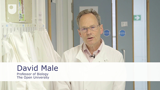
In the following video, David Male describes the subjects you will be covering this week, as you build on your ability to identify different types of cell.
Download this video clip.Video player: 39892_ou_futurelearn_mc1008_vid_008-540.mp4
Transcript
DAVID MALE
In the first part of the course, we showed you how to use a microscope, and how to recognise a variety of different cell types from blood under the microscope. In this part, we're going to introduce you to a variety of additional different types of human tissue. Often to help recognise structures within tissues, histologists use specialised stains on their sections. And we'll be showing you some examples of that, too. One of the ideas that I want to get over to you this week is that there is a range of normality within tissues.
So for example, the skin on my face, or the skin on my head, or the skin on my finger are all different, and will appear different underneath the microscope. But they are all quite normal. If, for example, I was a guitar player, I might have calluses on my fingers. But they would still be within the range of normality.
By the end of this week, you should be able to recognise a variety of different human tissues just from their histological appearance.
Interactive feature not available in single page view (see it in standard view).