Week 4: Recognising disease
Introduction
Welcome to the final week of the course.
Infection can affect any tissue of the body, producing cell damage and inflammatory reactions.
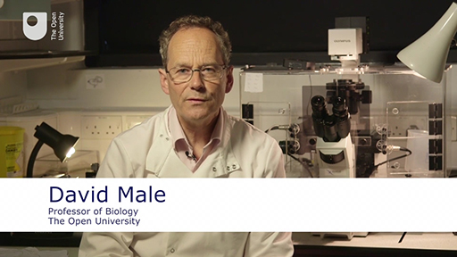
As David Male explains in the video below, the focus of this week is on studying pathological sections, namely those that show histological evidence of disease.

Transcript
The sections that you will look at have been organised around three main types of pathology:
- infection and inflammation
- degeneration and cell death
- tumours (specifically hyperplasia, dysplasia and neoplasia).
You will learn more about these topics as you progress through the week.
In the end-of-course quiz you’ll look at a number of different pathological sections to identify the abnormalities, to try to deduce the underlying causes and, finally, to venture a diagnosis of what the disease might be.
