

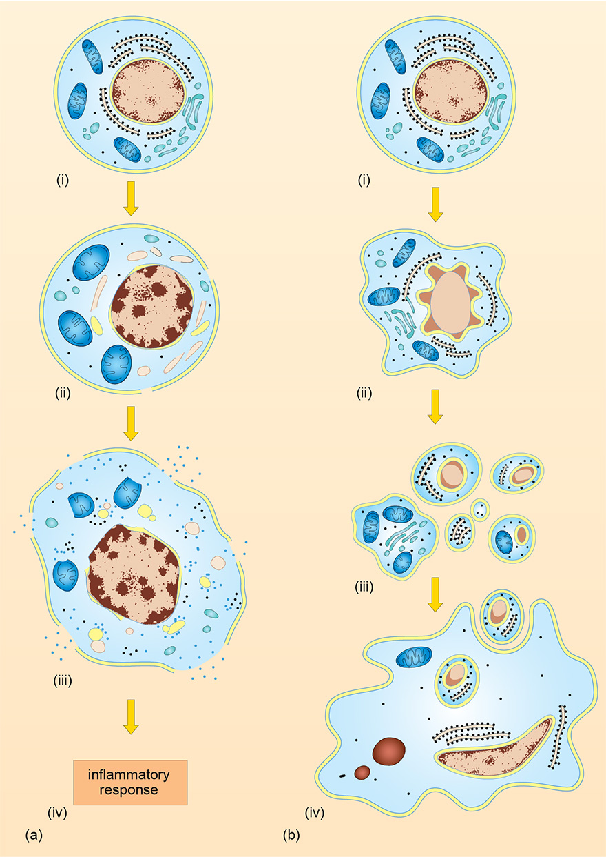
Figure 5 Schematic diagram comparing (a) necrosis and (b) apoptosis. The first events in necrosis are irregular condensation of the nucleus, swelling of the mitochondria and breakdown of membranes and ribosomes. The cell is eventually disrupted, releasing its contents and inducing an inflammatory reaction. In contrast, a cell undergoing apoptosis shows condensation of the nucleus into fragments and shrinkage of the cell. The nucleus and cytoplasm break up into fragments called apoptotic bodies, which are phagocytosed by mononuclear phagocytes.