

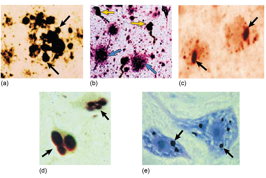
Figure 7 Protein aggregates in brain cells associated with neurodegenerative disease. Arrows highlight (a) extracellular plaques in prion disease, (b) extracellular plaques (blue) and neurofibrillary tangles (yellow) in Alzheimer’s disease, (c) nuclear aggregates in Huntington’s disease, (d) Lewy bodies in Parkinson’s disease, (e) nuclear aggregates in amyotrophic lateral sclerosis.