
Although a primary function of the inner ear is to perceive sound, it also houses the vestibular system. Information from the vestibular system is proprioceptive in character. This means that it deals with sensory events arising from within the body rather than exteroceptive events (sensory events that occur outside the body). Our sense of balance arises from the proprioceptive system as a whole which includes sensory receptors located within muscles and joints as well as those housed in the vestibular system. In this way balance of the whole body can be generated and monitored with the proprioceptors in the periphery (joints and muscles) ensuring and monitoring balance of our limbs, and the vestibular system ensuring and monitoring balance of our head. A functional vestibular system is important in everyday life and yet we are often only actively conscious of the vestibular system when it is damaged through illness. Such illnesses can be extremely debilitating, producing effects like vertigo (dizziness and loss of balance), which can considerably reduce a person’s quality of life.
The vestibular system is located in our inner ear. The inner ear consists of a system of tunnels: the bony labyrinth in the hardest part of the temporal bone. The two structures that make up the vestibular system – the vestibule and the semi-circular canals – are found within this labyrinth.
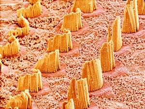
 The semicircular canals, part of the vestibular system, are shown here in white next to the spiral shaped cochlea.
The vestibule houses two membranous sacs – the utricle and the saccule – which are collectively known as otolithic organs. The semi-circular canals (there are three of them) are orientated at right angles to one another, and these are termed the superior, posterior and lateral canals. At each end of a canal there is a swelling of the tube called an ampulla and both ends of each canal open out into the vestibule. To give some impression of their size, the vestibule (associated with the otolithic organs) in humans is about 3-5mm across, while the semi-circular canals range from 12mm up to 22mm in length.
The semicircular canals, part of the vestibular system, are shown here in white next to the spiral shaped cochlea.
The vestibule houses two membranous sacs – the utricle and the saccule – which are collectively known as otolithic organs. The semi-circular canals (there are three of them) are orientated at right angles to one another, and these are termed the superior, posterior and lateral canals. At each end of a canal there is a swelling of the tube called an ampulla and both ends of each canal open out into the vestibule. To give some impression of their size, the vestibule (associated with the otolithic organs) in humans is about 3-5mm across, while the semi-circular canals range from 12mm up to 22mm in length.
The bony labyrinth that houses the otolithic organs and the semi-circular canals is lined by an outer membrane that encloses an inner membranous labyrinth. This arrangement means that the vestibular system contains two almost concentric compartments both filled with fluid. The outer compartment contains a type of fluid called perilymph and the inner compartment contains a fluid called endolymph. These two fluids vary in their concentrations of ions which are critical for maintaining the correct physiological state of cells.
The otolithic organs and the semi-circular canals contain hair cells which are the sensory receptors for the vestibular system. These hair cells are positioned within an epithelial layer that also contains the nerve processes connecting to the hair cells. Like all sensory systems, the nerve cells that connect to the sensory receptors (hair cells) ensure that the signals detected by the receptors can be sent to the brain for further perceptual processing.
The hair cells of both structures (the otolithic organs and the semi-circular canals) project into specific overlying structures. For the otolothic organs this is the otolithic membrane, the surface of which is covered by a blanket of crystals called otoliths. For the semi-circular canals this is a gelatinous mass called the cupula.
 There are two types of hair cells in the vestibular system. The first type are flask shaped and are innervated by a nerve cell that forms a cup around the cell body. This cup is sometimes shared by other hair cells. The second type of hair cells are more cylindrical and have small round nerve endings onto the cell body.
All hair cells are made up of hair like bundles of stereocilia and a single kinocilium. The stereocilia are composed of actin associated proteins like myosin which influence how they move while the kinocilium is built slightly differently. The stereocillia are arranged in order of size and the single kinocilium is larger than the longest stereocilia so it sits at the end of all the stereocilia. Actin proteins like myosin can control the tension in the stereocilia, which is how it affects the amount of movement they can exhibit. This is very important because the stereocilia move in response to turning or movement of the head. This occurs because, when the head moves the otoliths (crystals) in the utricle and saccule, and the endolymph (fluid) in the semi-circular canal lags behind due to inertia. The lag is communicated to the otolithic membrane (for the otoliths) and cupula (for the semicircular canals) respectively causing deflection of the hair cell bundles. In other words: this deflection is the sensory signal.
Deflection of the hair cells results in an electrical signal in the nerve cells connecting to them. This information is relayed in a bundle of these nerves, called the vestibular nerve, to the base of the brain where information from both ears and from the different components of the vestibular system (otolithic organ and semi-circular canals) is integrated. After this point the information ascends to be processed in the oculomotor system which is a system that coordinates eye movements in response to head movements. This process allows a stable image to be maintained on the retina of the eye when the head moves – a critical thing for maintaining balance. Thus visual information is also important for our balance. The stable retinal image is maintained by something called the vestibular-occular reflex which you can very easily demonstrate the effectiveness of for yourself. Simply shake your head while trying to read this article. You will find that you have no trouble staying focused on the text despite the erratic movement of your head!
Finally vestibular information enters a structure called the thalamus and from here it will be processed in the vestibular cortex. Interestingly there is still some debate about exactly where the vestibular cortex is located but it is thought to lie adjacent to the region of the cortex that processes information from the peripheral proprioceptive system (our muscles and joints). Recall from the start of this article that this system is responsible for balance of the body and works alongside the vestibular system which is responsible for balance of the head.
In sum therefore, how our sense of balance is created and maintained is complex, arising from a variety of systems in the body processing visual information, as well as peripheral proprioceptive and vestibular information. But we have many more sensory systems that ensure that we can respond appropriately to the world around us. Every single one of these sensory systems was carefully constructed during our time in the womb.
If you are interested in learning more about our sensory systems including how we perceive pain, taste, smell, hearing and vision, why not try our 3rd level module SD329 Signals and Perception: The Science of the Senses. This module can be studied as part of our degree in Health Sciences (Q71) as well as part of the Natural Sciences (Q64) and Open Degree.
It is, however, strongly recommended that you do not attempt a third level Open University module without prior Open University study at a lower level.
Explore more...
-
How testosterone affects risk taking behaviour
Read now to access more details of How testosterone affects risk taking behaviourWhat role does testosterone play in risk taking behaviour?
Level: 1 Introductory
Rate and Review
Rate this article
Review this article
Log into OpenLearn to leave reviews and join in the conversation.
Article reviews