

1.3 Staining techniques
Most cells and cellular elements are virtually transparent, so it is difficult to distinguish individual cells and cellular structures when viewed with a microscope. However, this issue can be countered if staining agents are applied to the sample while it is being made ready for analysis.
Histologists have developed a wide variety of different staining techniques to identify different elements within tissues when they are looked at with a microscope. This methodology is referred to as histochemistry.
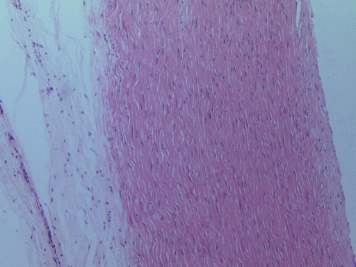
The most commonly used stain for medical diagnosis uses a combination of the dyes haematoxylin (which is blue) and eosin (which is red). This approach is usually abbreviated to ‘H&E’ staining. An example of some tissue stained using H&E is shown in the slide image below.
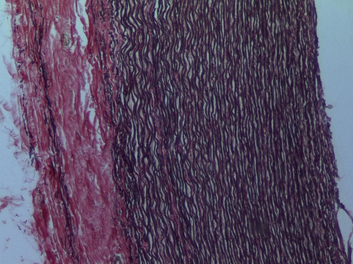
However, there are some structures that do not stain well with H&E, so different stains have been developed that allow the identification of particular tissue elements (e.g. collagen). These alternative stains include Giemsa, Masson’s trichrome and elastic Van Gieson (see the slide below, Figure 5), and they are described in more detail shortly.
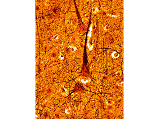
A wide variety of other histochemical stains are also available, each of which can help identify particular structures. Some are relatively simple to apply, merely requiring that the section is dipped in the stain for a set time. Others require a number of sequential steps, and in some cases the results can be surprisingly variable or unpredictable. For example, the silver staining technique, originally developed by Camillo Golgi and shown in Figure 6, is notably variable.
Some of the more commonly used staining techniques are outlined below.
- Giemsa stain consists of a mixture of methylene blue and eosin. It is mostly used on blood films, where it stains red blood cells (erythrocytes) pink and the different types of white blood cells (leukocytes), allowing their identification according to the size and shape of their nuclei. It also binds to some pathogens (e.g. spirochaetes and syphilis) and it is, therefore, particularly useful if infection is suspected.
- Luxol fast blue/cresyl violet is used to identify myelin, a component of the nervous system which insulates the nerves and stains blue, while other elements of the nervous system stain pink or violet.
- Masson’s trichrome stain uses three dyes to stain different structures. It is valuable for distinguishing elements of connective tissue. Typically the cell cytoplasm, muscle and keratin are stained red, nuclei are stained black and collagen is stained blue.
- Periodic acid schiff (pas) stains carbohydrates magenta, including components of the basal lamina, surface glycoproteins on cells, and intracellular carbohydrates, such as glycogen in hepatocytes. Cells that secrete mucus are also strongly stained.
- Congo red is used to identify deposits of protein in tissue called amyloid.
- Toluidine blue is a particularly versatile dye that stains nuclei blue, and can be used to differentiate between different types of granules (e.g. within mast cells).
- Van Gieson stain binds to collagen in the extracellular matrix, staining it pink. It is often combined with a stain for elastic fibres (elastic Van Gieson) which stains black, allowing the two major elements of connective tissue to be differentiated.