1.5 Cells and tissues
Cells have distinctive shapes and functions which depend on how they have developed and their interactions with other cell types. It is difficult to be precise, but there are probably at least 200 different types of cell within the body.
Disease processes affect individual cells in many ways. They may cause them to:
- die
- change their shape
- divide
- move
- invade other tissues.
Any of these changes also affect the anatomy of the tissue.
Understanding the changes that are characteristic of a disease requires a detailed knowledge of the normal appearance of cells and tissues, and the range of normality. Many tissues change considerably with age, so that a feature that is normal in an adult would not be normal in a child. For example, the thymus gland gradually decreases in size with age, so a large thymus in an old person could indicate some underlying pathology.
Primary and secondary changes
The histological changes seen in a tissue may be primary, a cause of the disease process, or secondary, a consequence.
For example, if blood pressure is high due to vascular disease, it may cause an increase in the volume of the heart muscle, as the heart finds it more difficult to pump blood into the circulation. A further consequence may be a corresponding decrease in the volume of the chambers of the heart so that less blood is pumped with each contraction.
Interpreting the changes seen histologically requires a sound understanding of the underlying disease processes.
Identifying cells and structures
As you have read, it is difficult to see many cellular structures using a light microscope, so dyes are often applied to samples to stain the contents and make them visible. There are two main ways of staining cells.
- The traditional method involves the use of dyes that selectively bind to different structures within the cell.
- More recently immunohistochemistry techniques have emerged that use antibodies to stain individual molecules.
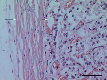
Most of the sections you will view in this course have been stained with haematoxylin and eosin (or H&E). The cell nucleus containing DNA binds haematoxylin and is, therefore, blue, as can be seen in Figure 8 below. Inactive cells usually stain dark blue, while more active cells are paler. Dead cells have shrunken compacted nuclei which may be fragmented.

The cytoplasm of a cell stained with H&E may range from pink to purple. More active cells have diffuse purple staining ribonucleic acid (RNA) in the cytoplasm indicating that they are producing protein. Many cells also have granules which are acidophilic (pink) or basophilic (purple) depending on whether they have taken up eosin or haematoxylin.
The cytoplasm may also contain non-staining vacuoles. For example, fat cells (adipocytes) have a large central vacuole containing fat.
