1.1 Recognising common cell types
This video describes how to use the virtual microscope tool to recognise some of the different cell structures that you have just read about.
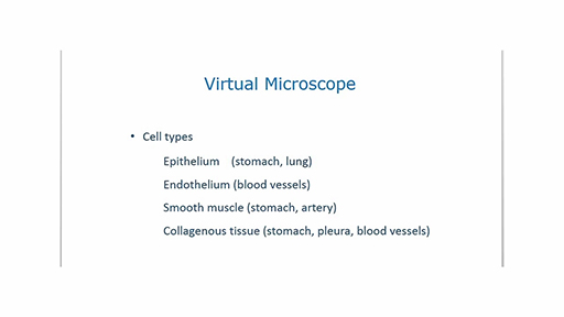
Download this video clip.Video player: 39448_ou_futurelearn_mc1008_vid_009-540.mp4
Transcript
PROFESSOR DAVID MALE
Cell types. In this video, I will look at some of the sections that you will be examining during the course. And I'll be looking for different types of cell. I'm not aiming to examine every detail of the sections. You'll have time to do that later. And the legends on the sections will help you to identify specific features.
I will be looking for different types of epithelium in the stomach and lung, endothelium in different blood vessels, smooth muscle in the wall of the stomach and an artery. And you will also see examples of collagenous tissue in different organs.
I'll start by looking at the section of stomach, which is slide number three. There is a layer of epithelial cells lining the inside of the stomach. If I look on the times 20 magnification, you can see that this is a single layer of columnar epithelium.
Because of the way a section is cut, the appearance of the cells may not always correspond to the classical columnar form, as you can see here. And if I move across, also here.
If I move to the next layer of cells, the submucosa, and I return to the times 20 magnification, you'll see an example of collagenous tissue. There are relatively few cells and a large amount of extracellular space, of which a large proportion is collagen secreted by the cells in this layer.
Further out again is a layer of smooth muscle cells. These cells are densely packed. And they have elongated nuclei. And they run in lines corresponding to the direction of contraction of the muscle. The smooth muscle in the gut propels food through the system.
I'm now going to look at a blood vessel, in this case, the cross-section of a small artery. There's a single layer of endothelial cells lining the inside of this vessel. The cells and their nuclei are very thin and flattened.
The artery also has smooth muscle in its wall, which resists the pressure from the blood flow and partly controls the amount of blood that can pass through the lumen of the vessel. Around the edge of the vessel, there is a layer of collagenous tissue referred to as the adventitia.
Finally, I'm going to look at a section of lung. The outside of the lung has an outer surface called the pleura. It consists primarily of collagenous tissue, which you can see at the high magnification. The main cell types in the lung are epithelial cells called pneumocytes, which are involved in gas exchange. There are some examples here. There are also many capillary blood vessels and small arteries called arterials.
As you start to explore the sections, you will find common patterns of cellular organisation in different tissues and organs.
Interactive feature not available in single page view (see it in standard view).
You will explore some of these cell types in more detail as you progress through the rest of the course. However, what is important to recognise here is the fact that these distinct structures commonly recur in very different types of body tissue.